Quick Facts
Origin: Posterior surface of tibia, inferior to soleal line, posterior surface of fibula, and adjacent interosseous membrane of leg.
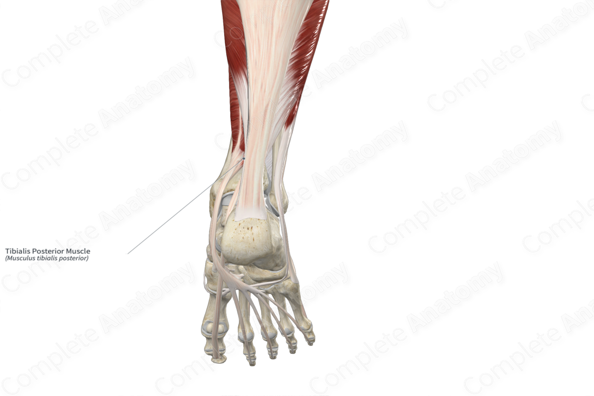
Insertion: Tuberosity of navicular bone, plantar aspects of medial, intermediate, and lateral cuneiform bones, and plantar aspects of bases of second to fourth metatarsal bones.
Action: Inverts foot at subtalar and transverse tarsal joints; plantarflexes foot at ankle joint.
Innervation: Tibial nerve (L4-L5).
Arterial Supply: Posterior tibial and fibular arteries.


Related parts of the anatomy
Origin
The tibialis posterior muscle originates from the:
- lateral aspect of the area of the posterior surface of the tibia that is located inferior to the soleal line;
- medial aspect of the proximal two thirds of the posterior surface of the fibula;
- posterior aspect of the adjacent interosseous membrane of leg;
- adjacent intermuscular septa.
Insertion
The fibers of the tibialis posterior muscle travel inferiorly to the foot and insert, via a long tendon, onto the:
- tuberosity of navicular bone;
- plantar aspects of the medial, intermediate and lateral cuneiform bones;
- plantar aspects of the bases of the second to fourth metatarsal bones.
In some individuals, the tibialis posterior muscle can also insert onto the sustentaculum tali of calcaneus and the plantar aspect of the cuboid bone.
Key Features & Anatomical Relations
The tibialis posterior muscle is one of the muscles of the deep part of the posterior compartment of the leg. It is a long, narrow, unipennate type of skeletal muscle. In the distal one third of the leg, the muscle belly gives rise to a tendon that travels inferomedially towards the malleolar groove of the tibia. The tendon hooks around the groove, changing its line of pull to a more anteroinferior direction. This tendon then travels deep to the flexor retinaculum of foot, anterior to the adjacent tendon of flexor digitorum longus, where it passes through the tendinous sheath of tibialis posterior muscle.
Along the lateral aspect of the foot, the tendon continues to travel anteroinferiorly, passing medial to the medial collateral ligament of the ankle joint, then inferomedial to the plantar calcaneonavicular ligament and then to its insertion sites.
Within the posterior compartment of the leg, the tibialis posterior muscle is located:
- anterior to the transverse intermuscular septum of leg, the flexor hallucis longus and soleus muscles, the posterior tibial vessels, and the tibial nerve;
- posterior to the tibia, fibula and interosseous membrane of leg;
- lateral to the flexor digitorum longus muscle.
Within the plantar part of the foot:
- the tendon of the tibialis posterior muscle is located deep to the abductor hallucis muscle and the tendon of the flexor digitorum longus;
- the medial of the two proximal tendons of the flexor hallucis brevis muscle originates from the tendon of the tibialis posterior.
The tibialis posterior muscle is one of five structures that pass posterior to the medial malleolus. From anterior to posterior, these are the tibialis posterior muscle, flexor digitorum longus muscle, posterior tibial artery, tibial nerve, and flexor hallucis longus muscle.
Actions & Testing
The tibialis posterior muscle is involved in multiple actions:
- inverts the foot at the subtalar and transverse tarsal joints;
- plantarflexes the foot at the ankle joint;
- helps stabilize the longitudinal arch of the foot (Sinnatamby, 2011).
The tibialis posterior muscle can be tested by inverting the foot against resistance while the foot is held in the plantarflexed position, during which its tendon can be seen and palpated (Standring, 2016).
List of Clinical Correlates
- Posterior shin splints
- Pes planus
References
Sinnatamby, C. S. (2011) Last's Anatomy: Regional and Applied. ClinicalKey 2012: Churchill Livingstone/Elsevier.
Standring, S. (2016) Gray's Anatomy: The Anatomical Basis of Clinical Practice. Gray's Anatomy Series 41st edn.: Elsevier Limited.
Learn more about this topic from other Elsevier products

