Quick Facts
Origin: Digastric fossa of mandible.
Insertion: Intermediate tendon of digastric muscle.
Action: Depresses mandible and elevates hyoid bone.
Innervation: Digastric branch of nerve to mylohyoid muscle (CN V3).
Arterial Supply: Submental artery.


Origin
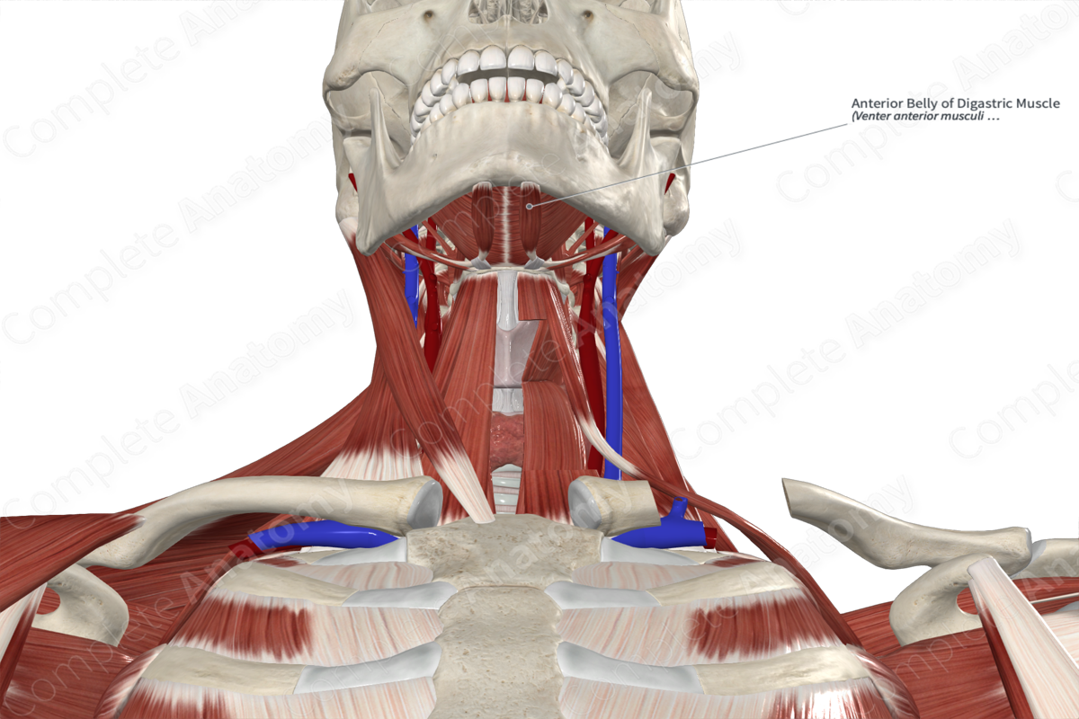
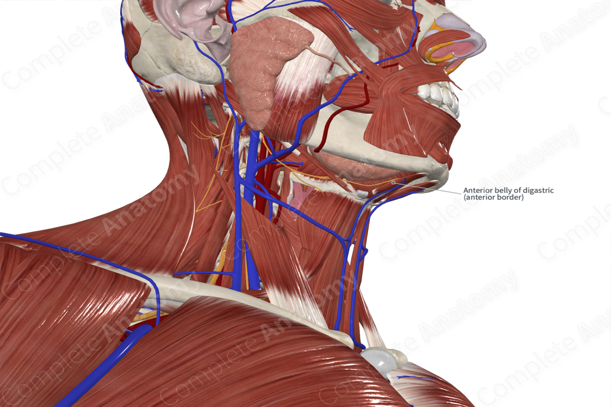
The anterior belly of digastric muscle originates from the digastric fossa, which is found along the base of the mandible.
Insertion
Both the anterior and posterior bellies of the digastric muscle insert into the intermediate tendon of digastric muscle. This tendon pierces the stylohyoid muscle and then passes through a fibrous sling that is attached to the hyoid bone.
The fibers of the anterior belly run on the external surface of mylohyoid muscle before attaching to the intermediate tendon.
Key Features & Anatomical Relations
The anterior and posterior bellies of the digastric muscle divide the anterior triangle of the neck into submandibular and sublingual triangles. The anterior belly of digastric sits superficial to the mylohyoid muscle.
The fact that the anterior and posterior bellies have separate innervation reflects their embryological origins. The anterior belly is derived from the first pharyngeal arch and, thus, is innervated by the trigeminal nerve (CN V). The posterior belly of the digastric muscle is derived from the second pharyngeal arch and, therefore, it is innervated by the facial nerve (CN VII).
Actions
Overall, the digastric muscle is involved in multiple actions:
- depresses the mandible at the temporomandibular joint;
- elevates the hyoid bone (Standring, 2016).
References
Standring, S. (2016) Gray's Anatomy: The Anatomical Basis of Clinical Practice. Gray's Anatomy Series 41st edn.: Elsevier Limited.
