Quick Facts
Origin: Posterior belly: mastoid notch of temporal bone; Anterior belly: digastric fossa of mandible.
Insertion: Intermediate tendon of digastric muscle.
Action: Depresses mandible; elevates hyoid bone.
Innervation: Posterior belly: digastric branch of facial nerve (CN VII); Anterior belly: digastric branch of nerve to mylohyoid muscle (CN V3).
Arterial Supply: Posterior belly: posterior auricular and occipital arteries; Anterior belly: submental artery.


Origin
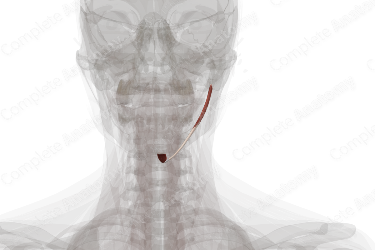
The digastric muscle, as its name implies, has two muscular bellies. These are the anterior and posterior bellies and are connected by a small intermediate tendon.
The posterior belly originates from the mastoid notch, which is found along the internal surface of the mastoid process of temporal bone. The anterior belly originates from the digastric fossa, which is found along the base of the mandible.
Insertion
Both the anterior and posterior bellies of the digastric muscle insert into the intermediate tendon of digastric muscle. This tendon pierces the stylohyoid muscle and then passes through a fibrous sling that is attached to the hyoid bone.
The fibers of the posterior belly descend anteriorly before attaching to the intermediate tendon, while the fibers of the anterior belly run on the external surface of mylohyoid muscle before attaching to the intermediate tendon.
Key Features & Anatomical Relations
The anterior and posterior bellies of the digastric muscle divide the anterior triangle of the neck into submandibular and sublingual triangles.
The platysma, sternocleidomastoid, stylohyoid, and longissimus capitis muscles, as well as the mastoid process of the temporal bone, sit superficial to the digastric muscle. The retromandibular vein and the submandibular and parotid glands lie over the digastric muscle. The anterior belly of digastric sits superficial to the mylohyoid muscle.
The fact that the anterior and posterior bellies have separate innervation reflects their embryological origins. The anterior belly is derived from the first pharyngeal arch and, thus, is innervated by the trigeminal nerve (CN V). The posterior belly of the digastric muscle is derived from the second pharyngeal arch and, therefore, it is innervated by the facial nerve (CN VII).
Actions
Overall, the digastric muscle is involved in multiple actions:
- depresses the mandible at the temporomandibular joint;
- elevates the hyoid bone (Standring, 2016).
References
Standring, S. (2016) Gray's Anatomy: The Anatomical Basis of Clinical Practice. Gray's Anatomy Series 41st edn.: Elsevier Limited.
Learn more about this topic from other Elsevier products

