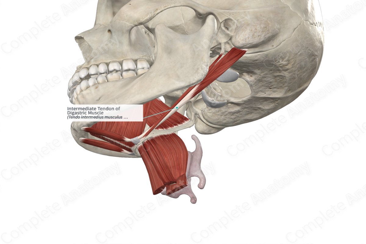
Intermediate Tendon of Digastric Muscle
Tendo intermedius musculus digastrici
Read moreStructure/Morphology
The intermediate tendon of the digastric muscle connects the anterior and posterior bellies of the digastric muscle.


Anatomical Relations
The intermediate tendon of the digastric muscle is closely associated with the hyoid bone as it passed below a fibrous loop on its anterior surface. The posterior belly, or the intermediate tendon, penetrates the stylohyoid muscle as it approaches its insertion on the hyoid bone (Kim and Loukas, 2019).
In some instances, the tendon is varied, the most common of which is that it gives off two anterior bellies as well as its normal posterior belly (Kim and Loukas, 2019).
Function
Since the intermediate tendon of the digastric muscle is attached to the hyoid bone via a fibrous loop, contraction of the digastric muscle causes elevation of the hyoid bone. If the hyoid bone is fixed by contraction of the infrahyoid, contraction of digastric muscle will cause depression of the mandible (Netter, 2011).
References
Kim, S. D. and Loukas, M. (2019) 'Anatomy and variations of digastric muscle', Anatomy & cell biology, 52(1), pp. 1-11.
Netter, F. H. (2011) Atlas of Human Anatomy. Netter Basic Science Series: Saunders/Elsevier.
Learn more about this topic from other Elsevier products

