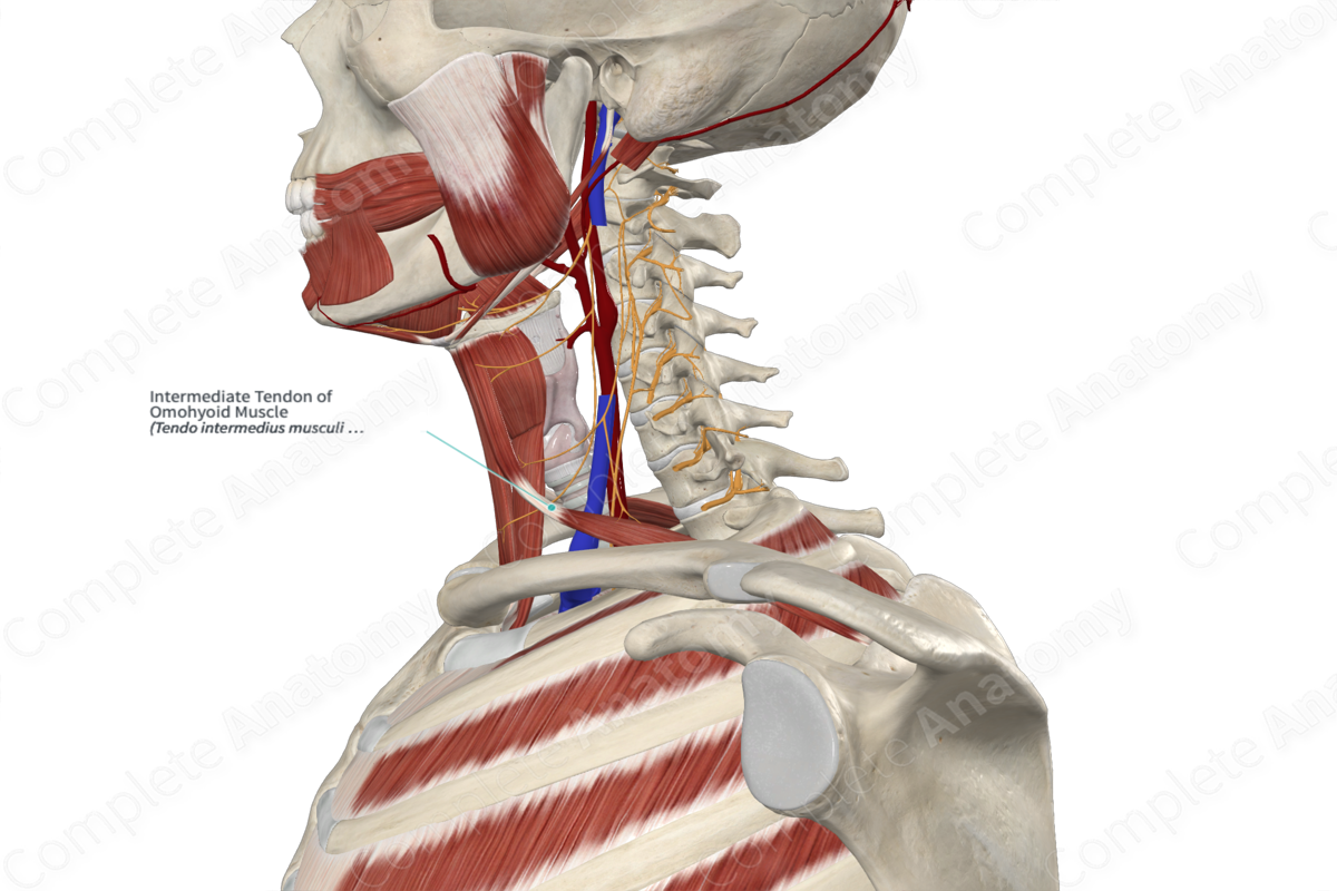
Intermediate Tendon of Omohyoid Muscle
Tendo intermedius musculi omohyoidei
Read moreStructure/Morphology
The omohyoid muscle is made up of two bellies united at an angle by an intermediate tendon. A band of deep cervical fascia, attached to the superior border of the body of the clavicle houses the intermediate tendon and maintains the angulated course of the muscle (Standring, 2016).


Related parts of the anatomy
Anatomical Relations
The intermediate tendon of the omohyoid muscle lies adjacent to the internal jugular vein at the level of the arch of the cricoid cartilage.
Function
The intermediate tendon of the omohyoid muscle connects the anterior and posterior bellies of omohyoid muscle, of which the function is to depress the hyoid bone.
References
Standring, S. (2016) Gray's Anatomy: The Anatomical Basis of Clinical Practice. Gray's Anatomy Series 41st edn.: Elsevier Limited.
