Quick Facts
Origin: Greater and lesser horns of hyoid bone; stylohyoid ligament.
Insertion: Pharyngeal raphe.
Action: Constriction of mid-pharynx during swallowing.
Innervation: Pharyngeal plexus (CN X).
Arterial Supply: Ascending pharyngeal artery and tonsillar branch of facial artery.


Origin
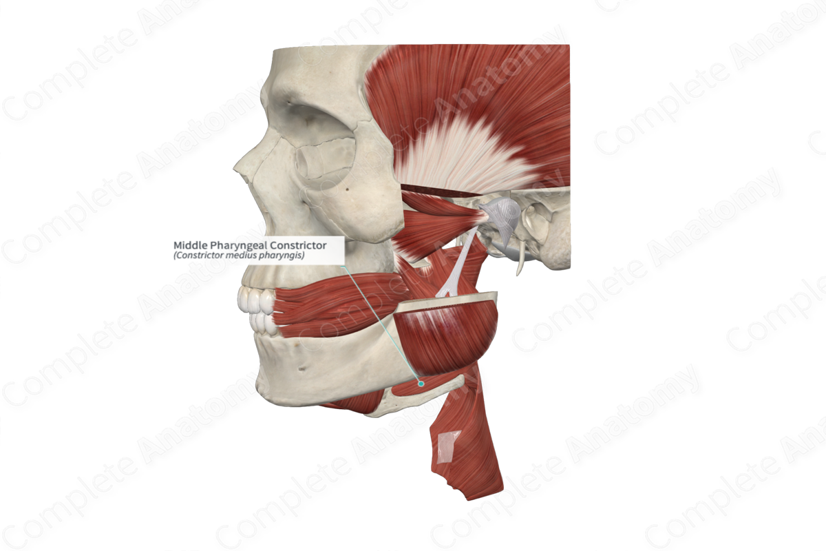
The middle pharyngeal constrictor muscle originates from the greater and lesser horns of the hyoid bone and the inferior region of the stylohyoid ligament.
Insertion
The fibers of the middle pharyngeal constrictor muscle insert into the pharyngeal raphe on the posterior aspect of the pharynx.
Key Features & Anatomical Relations
The three pharyngeal constrictor muscles overlap whereby the lower border of the superior constrictor sits deep to the middle constrictor and, in turn, the lower border of the middle constrictor sits deep to the inferior constrictor muscle (i.e., like a stack of cups).
The middle and superior constrictor muscles are separated by stylopharyngeus and the glossopharyngeal nerve (CN IX). Additionally, the palatopharyngeus muscle sits internal to the middle pharyngeal constrictor muscle.
Actions
The middle pharyngeal constrictor muscle constricts the middle part of the pharynx during swallowing (Standring, 2016).
References
Standring, S. (2016) Gray's Anatomy: The Anatomical Basis of Clinical Practice. Gray's Anatomy Series 41st edn.: Elsevier Limited.
