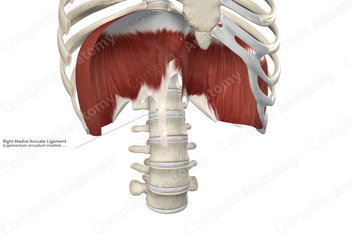
Structure/Morphology
The medial arcuate ligament is the arch-like, thickened area of the fascia covering the psoas major muscle. It attaches to the transverse process of the first lumbar vertebra, extends anteromedially across the anterior aspect of the psoas major muscle, and attaches to the anterolateral aspects of the vertebral bodies of the first and second lumbar vertebrae.
Anatomical Relations
There are five arcuate ligaments associated with the diaphragm:
- one median arcuate ligament;
- two medial arcuate ligaments;
- two lateral arcuate ligaments.
The medial arcuate ligament is located:
- anterior to the psoas major muscle;
- lateral to the first and second lumbar vertebrae and the crura of the diaphragm.
The sympathetic trunk passes through the gap formed by the medial arcuate ligament.
Function
Both the right and left medial arcuate ligaments provide attachment sites for some of the fibers of the lumbar part of the diaphragm.
Learn more about this topic from other Elsevier products
Joint Ligament

Entheseal structures are widely located throughout the body and are represented by the interface between bone and several tissues including tendon, joint capsules and ligaments.


