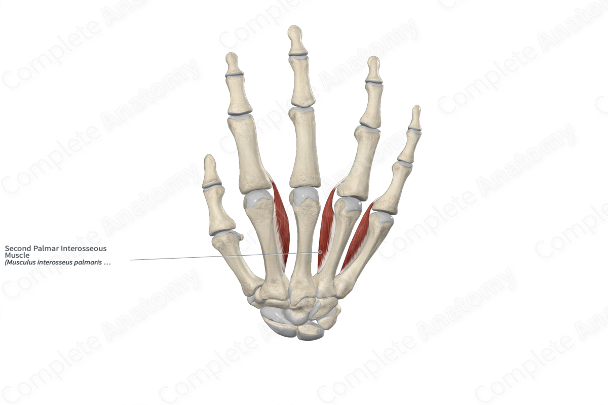
Quick Facts
Origin: Palmar aspect of fourth metacarpal bone.
Insertion: Lateral aspect of base of proximal phalanx and extensor expansion of ring finger.
Action: Adducts ring finger at its metacarpophalangeal joint; simultaneously flexes metacarpophalangeal joint and extends interphalangeal joints of ring finger.
Innervation: Deep branch of ulnar nerve (C8-T1).
Arterial Supply: Deep palmar arch, perforating branches of deep palmar arch, palmar metacarpal and common palmar digital arteries.
Related parts of the anatomy
Origin
The second palmar interosseous muscle originates from the palmar aspect of fourth metacarpal bone.
Insertion
The fibers of the second palmar interosseous muscle travel inferiorly to the ring finger and insert, via a short tendon, onto the:
- lateral aspect of the base of the proximal phalanx of ring finger;
- extensor expansion of ring finger.
Key Features & Anatomical Relations
The second palmar interosseous muscle is found in the interosseous compartment of the hand. It is a short, unipennate skeletal muscle. It is located:
- anterior to the fourth metacarpal bone and the third dorsal interosseous muscle of hand;
- posterior to the third lumbrical muscle of hand.
Actions & Testing
The second palmar interosseous muscle is involved in multiple actions:
- adducts the proximal phalanx of the ring finger (i.e., draws it towards the longitudinal axial line of the middle finger) at the fourth metacarpophalangeal joint;
- simultaneously flexes the fourth metacarpophalangeal joint and extends the interphalangeal joints of the ring finger, which occurs when the third lumbrical and fourth dorsal interosseous muscles of hand contract simultaneously with it.
The second palmar interosseous muscle can be tested by adducting the proximal phalanx of the ring finger at the fourth metacarpophalangeal joint against resistance (Standring, 2016).
References
Standring, S. (2016) Gray's Anatomy: The Anatomical Basis of Clinical Practice. Gray's Anatomy Series 41st edn.: Elsevier Limited.
Learn more about this topic from other Elsevier products
Hand Muscle

39,40 The lumbricals are a group of intrinsic hand muscles that originate on the flexor digitorum profundus (FDP) tendons.


