Quick Facts
Origin: Posterior aspect of body of pubis and tendinous arch of levator ani.
Insertion: Coccyx, pubococcygeus tendon, perineal body, and lateral walls of vagina in females and prostate in males.
Action: Provides structural support to adjacent pelvic structures; urinary and fecal continence.
Innervation: Nerve to levator ani muscle (S3-S4); inferior anal and perineal nerves.
Arterial Supply: Inferior gluteal, inferior vesical, and internal pudendal arteries.


Related parts of the anatomy
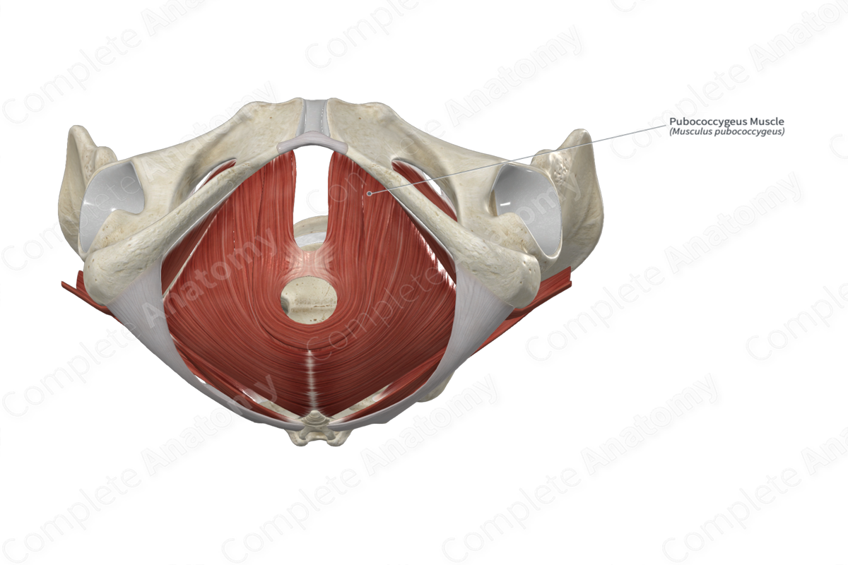
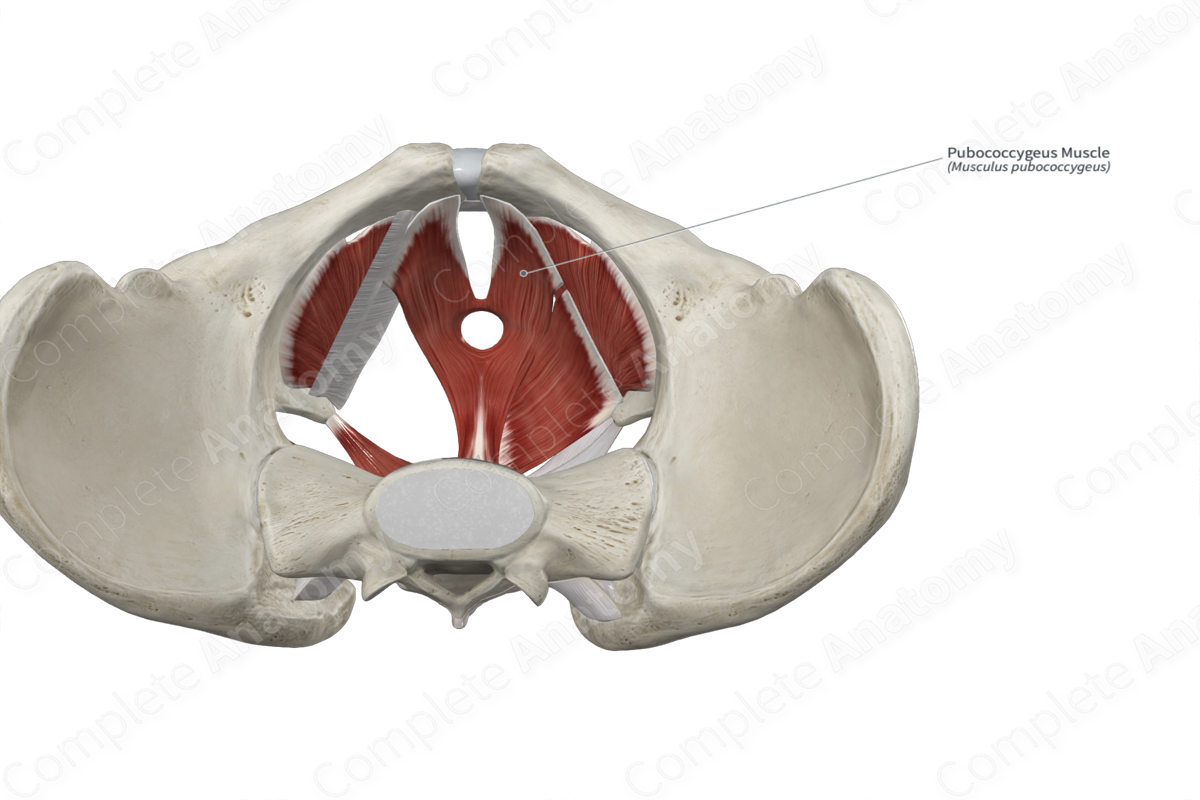
Origin
The pubococcygeus muscle originates from the:
- posterior aspect of the body of pubis;
- anterior portion of the tendinous arch of levator ani.
Insertion
The fibers of the pubococcygeus muscle travel posteriorly and can be arbitrarily divided into lateral and medial fibers.
- The lateral fibers of the pubococcygeus muscle insert onto the pelvic surface of coccyx and the pubococcygeus tendon. The pubococcygeus tendon is the fibrous, midline intersection between the lateral fibers of both the left and right pubococcygeus muscles. This tendon blends with the anococcygeal ligament.
- The medial fibers of the pubococcygeus muscle insert onto the lateral walls of the vagina in females and the lateral walls of the prostate in males. In both sexes, the left and right pubococcygeus muscles insert onto the perineal body and become continuous with each other as they arch over the posterior aspect of the urethra.
Key Features & Anatomical Relations
The pubococcygeus muscle is one of the three muscles that form the levator ani muscle, which itself forms a large part of the pelvic diaphragm. The pubococcygeus is a broad, flat skeletal muscle. It is located:
- superior to the puboanalis muscle;
- medial to the pubis and the iliococcygeus muscle;
- lateral to the urethra, the rectum, the vagina in females, and the prostate in males.
Some fibers of the pubococcygeus muscle have specific names:
- the fibers that lie adjacent to and attach to the vagina in females are known as the pubovaginalis muscle:
- the fibers that lie adjacent to and attach to the prostate in males are known as the puboprostaticus muscle.
Actions
As part of the pelvic diaphragm, the pubococcygeus muscle provides structural support to adjacent pelvic structures and elevates the pelvic floor. Its fibers are capable of maintaining a tonic contraction at rest, which relaxes during micturition and defecation (Standring, 2016).
List of Clinical Correlates
- Prolapse of pelvic viscera
- Urinary incontinence
- Fecal incontinence
References
Standring, S. (2016) Gray's Anatomy: The Anatomical Basis of Clinical Practice. Gray's Anatomy Series 41st edn.: Elsevier Limited.
Actions
As part of the pelvic diaphragm, the pubococcygeus muscle provides structural support to adjacent pelvic structures and elevates the pelvic floor. Its fibers are capable of maintaining a tonic contraction at rest, which relaxes during micturition and defecation (Standring, 2016).
