Quick Facts
Origin: Rami of both left and right ischial bones.
Insertion: Fibers surround anterior aspect of urethra.
Action: Constricts urethra.
Innervation: Perineal nerve (S2-S4).
Arterial Supply: Perineal artery.


Related parts of the anatomy
Origin
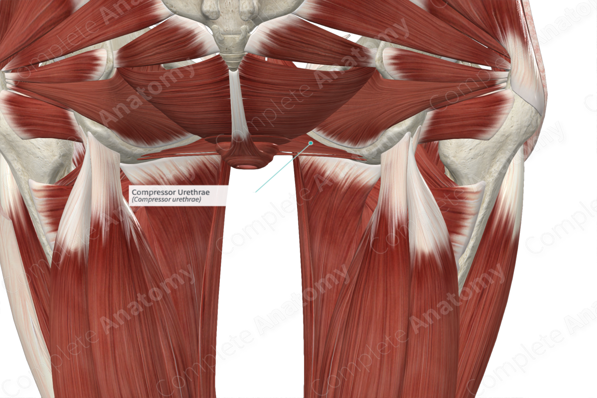
The compressor urethrae muscle originates from the rami of both the left and right ischial bones.
Insertion
The fibers of the compressor urethrae muscle travel medially, from both its left and right origin sites, to the anterior aspect of the urethra. Its left- and right-sided fibers are continuous with each other as they arch over the anterior aspect of the urethra (Jung, Ahn and Huh, 2012).
Key Features & Anatomical Relations
The female external urethral sphincter muscle is one of the muscles of the deep perineal space, which itself is found in the urogenital triangle. It is a large, circular muscle that partly surrounds both the urethra and vagina in females.
It is located:
- anterior to the perineal body;
- superior to the perineal membrane;
- inferior to the urinary bladder.
For descriptive purposes, the female external urethral sphincter muscle is divided into three parts:
- a superiorly located sphincter urethrae muscle;
- an anteriorly located compressor urethrae muscle;
- a posteriorly located sphincter urethrovaginalis muscle (Jung, Ahn and Huh, 2012).
Regarding the compressor urethrae muscle specifically, it is continuous:
- superiorly, with the sphincter urethrae muscle;
- posteriorly, with the sphincter urethrovaginalis muscle.
Actions
The female external urethral sphincter muscle constricts the urethra, keeping it closed and contributing to urinary continence. Its fibers relax during micturition (Standring, 2016).
List of Clinical Correlates
- Urinary incontinence
References
Jung, J., Ahn, H. K. and Huh, Y. (2012) 'Clinical and functional anatomy of the urethral sphincter', Int Neurourol J, 16(3), pp. 102-6.
Standring, S. (2016) Gray's Anatomy: The Anatomical Basis of Clinical Practice. Gray's Anatomy Series 41st edn.: Elsevier Limited.
