External Anal Sphincter Quick Facts
Origin: Anococcygeal ligament.
Insertion: Perineal body.
Action: Constricts anal canal.
Innervation: Perineal and inferior anal nerves.
Arterial Supply: Inferior anorectal artery.


External Anal Sphincter Origin
The external anal sphincter muscle originates from the anococcygeal ligament, which attaches to the apex of coccyx.
External Anal Sphincter Insertion
The fibers of the external anal sphincter muscle travel anteriorly and insert onto the perineal body.
External Anal Sphincter Key Features & Anatomical Relations
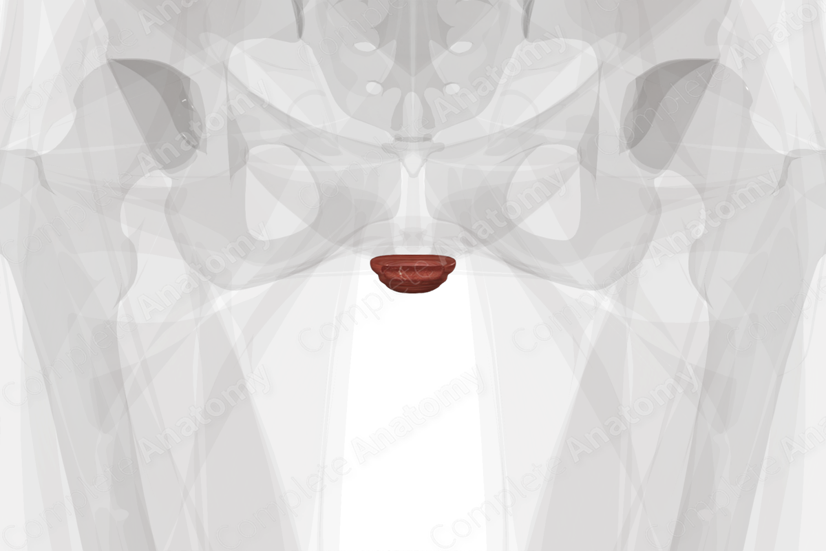
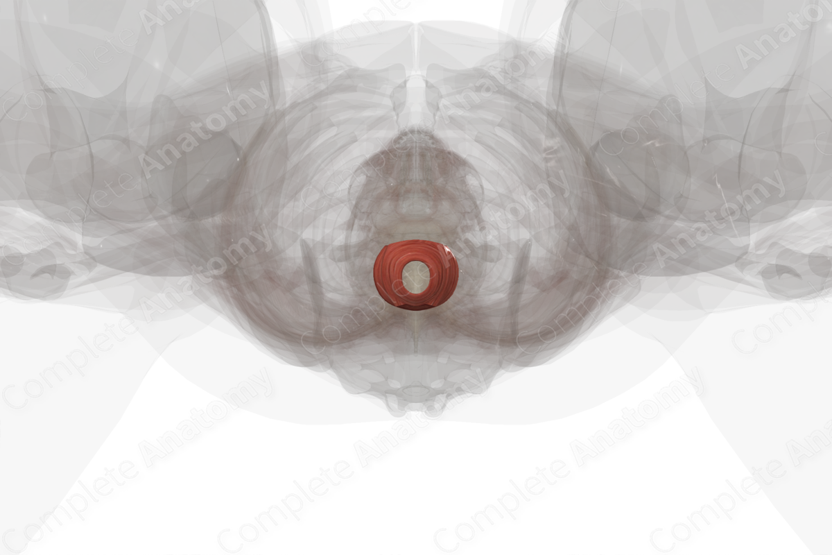
The external anal sphincter muscle is found in the anal triangle of the perineum. It is a large, voluntary, circular type of skeletal muscle that surrounds the anal canal.
For descriptive purposes, it is divided into three parts:
- a superiorly located deep part;
- a centrally located superficial part;
- an inferiorly located subcutaneous part.
The external anal sphincter muscle is located:
- anterior to the anococcygeal ligament;
- posterior to the perineal body and the superficial and deep transverse perineal muscles;
- inferior to the levator ani muscle.
External Anal Sphincter Actions
The external anal sphincter muscle constricts the anal canal, keeping it closed and contributing to fecal continence. Its fibers are capable of maintaining a tonic contraction at rest, which relaxes during defecation (Moore, Dalley and Agur, 2009).
External Anal Sphincter List of Clinical Correlates
- Fecal incontinence
External Anal Sphincter References
Moore, K. L., Dalley, A. F. and Agur, A. M. R. (2009) Clinically Oriented Anatomy. Lippincott Williams & Wilkins.
External Anal Sphincter Actions
The external anal sphincter muscle constricts the anal canal, keeping it closed and contributing to fecal continence. Its fibers are capable of maintaining a tonic contraction at rest, which relaxes during defecation (Moore, Dalley and Agur, 2009).
