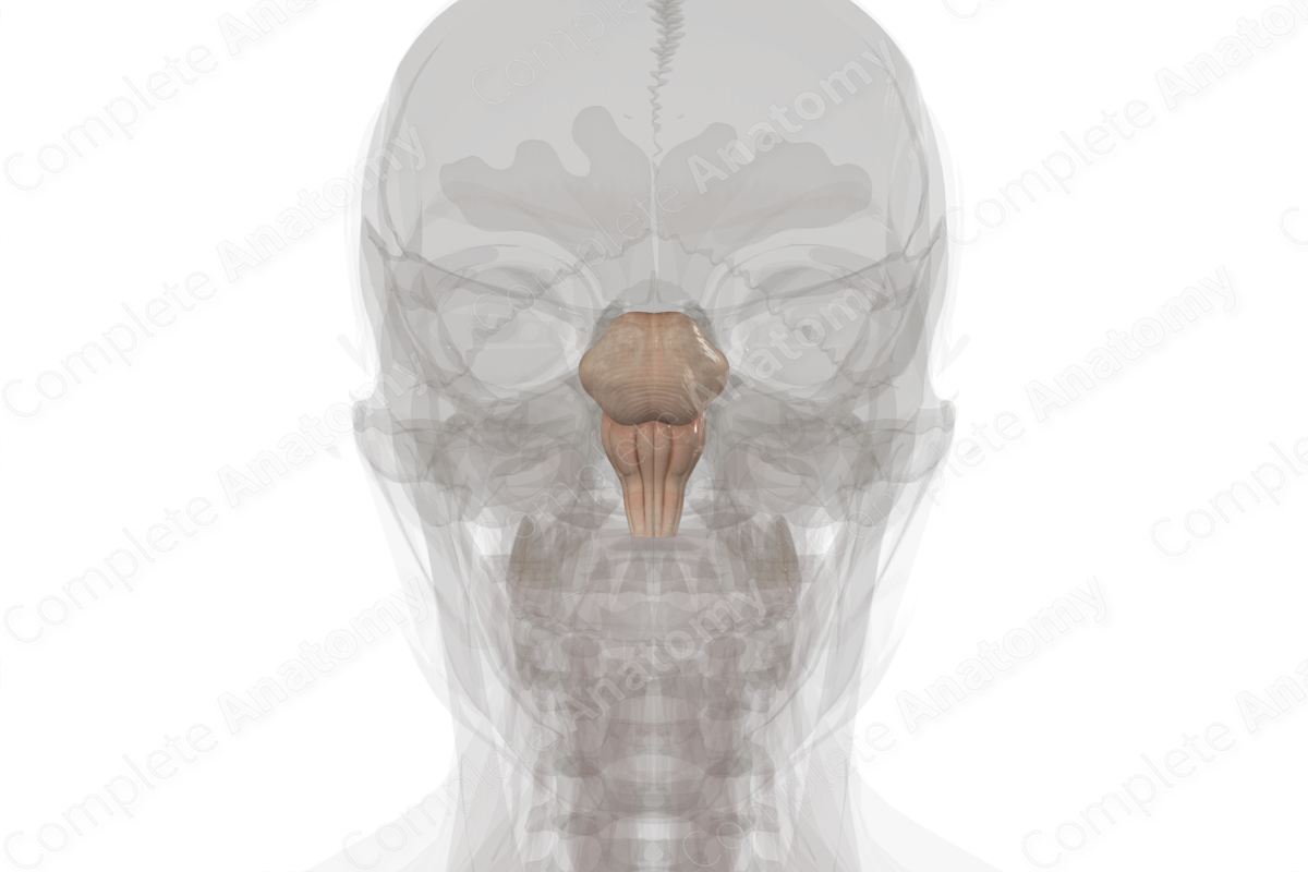
Quick Facts
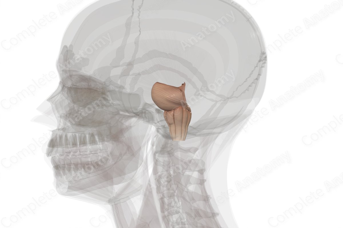
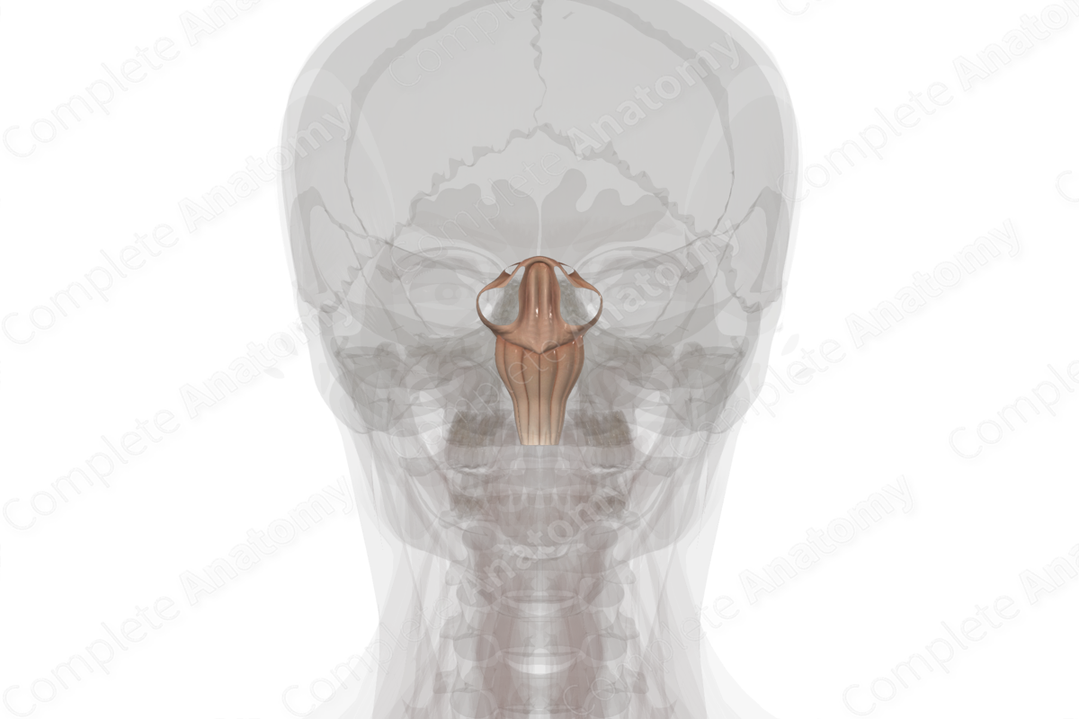
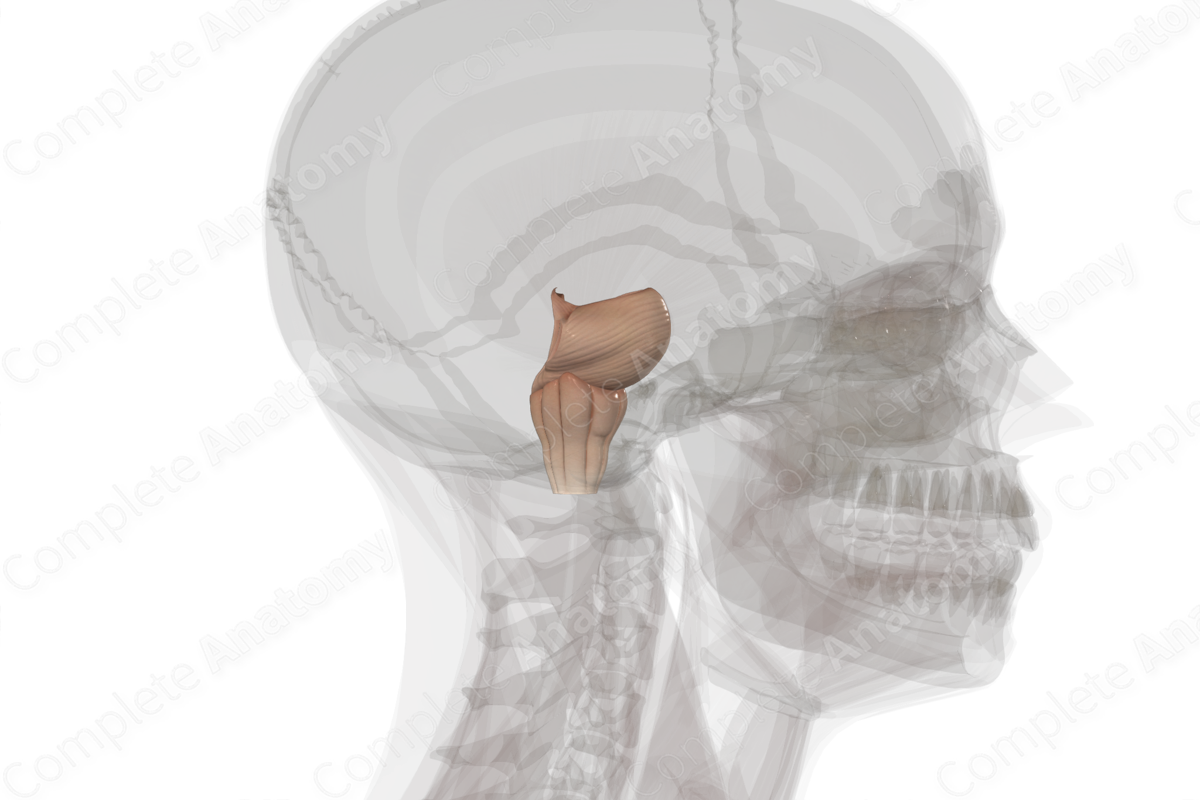
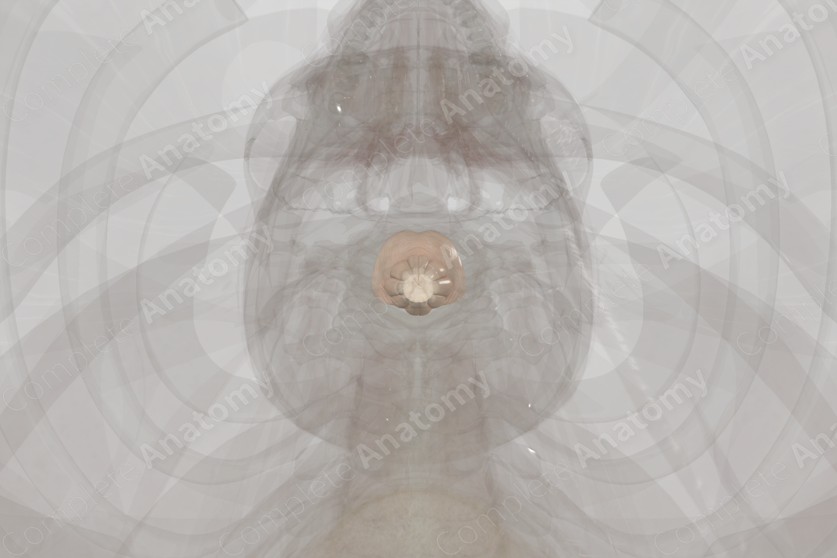
The brainstem is formed by the midbrain, the pons and the medulla oblongata and it connects the spinal cord with the brain. As well as being the site of origin of the optic and olfactory nerves, the brainstem is the part of the brain from which the cranial nerves emerge to provide the face and neck with motor and sensory innervation.
The descending motor signals from the cerebrum and cerebellum, and the ascending sensory signals from the body all pass through motor and sensory tracts in the brainstem. The motor signals from the cerebral cortex to the body are transmitted through the pyramidal tracts in the brainstem.
These pyramidal tracts can be divided into corticospinal tracts, which transmit signals to the spinal cord, and corticobulbar tracts, which transmit signals to the cranial nerves. The sensory signals arriving at the brain from the spinal cord travel through the posterior column-medial lemniscus pathway (PCML) in the brainstem.
The gracile and cuneate tubercles are part of the posterior column-medial lemniscus pathway. The cuneate nucleus, which forms the cuneate tubercle, carries fine touch and proprioceptive sensory information to the brain from the upper part of the body (above T6), while the gracile nucleus that makes up the gracile tubercle carries sensory information to the brain from the lower part of the body (below T6).
The pons is a bulbous protrusion, located above the medulla oblongata and below the midbrain. It contains white matter tracts, which transmit motor signals from the cerebral cortex to the cerebellum and spinal cord and sensory signals from the spinal cord to the thalamus. It also contains a number of nuclei involved with balance, posture, facial sensation and facial muscle control, sleep and respiration.
Related parts of the anatomy
Learn more about this topic from other Elsevier products
Brain Stem

The brain stem is generally a solid cylinder of neural tissue, both white and gray matter, found between the diencephalon and spinal cord and appears to play a central role in the linkage of the spinal cord (and thereby the body) and nonneural cranium (via the cranial nerves) with the regions of the brain involved in higher order processing (diencephalon and telencephalon).

