Structure
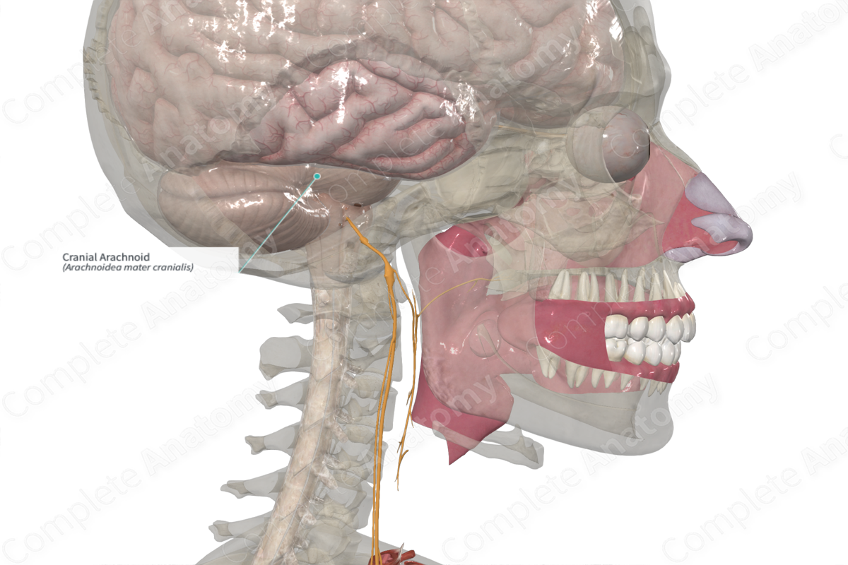
The arachnoid mater is the middle of the three meningeal layers. It is a thin opaque layer that sends wispy fibers inward to the pia mater, forming the spider web appearance that gives the arachnoid mater its name. Where it overlies a dural venous sinus, the arachnoid mater can develop arachnoid granulations, extensions of the arachnoid that pierce the dura and allow cerebrospinal fluid to flow out of the subarachnoid space and into venous sinuses. The cranial arachnoid mater is similar in function and appearance to the spinal arachnoid mater.


Related parts of the anatomy
Anatomical Relations
The arachnoid mater lies against the dura mater, being held away from the brain and pia mater by the presence of cerebrospinal fluid. This space found between the pia and the arachnoid mater is the subarachnoid space and contains cerebrospinal fluid.
List of Clinical Correlates
- Subarachnoid hemorrhage
Learn more about this topic from other Elsevier products
Arachnoid

A SAH is a type of haemorrhagic stroke characterised by bleeding into the subarachnoid space around the brain.
