Structure
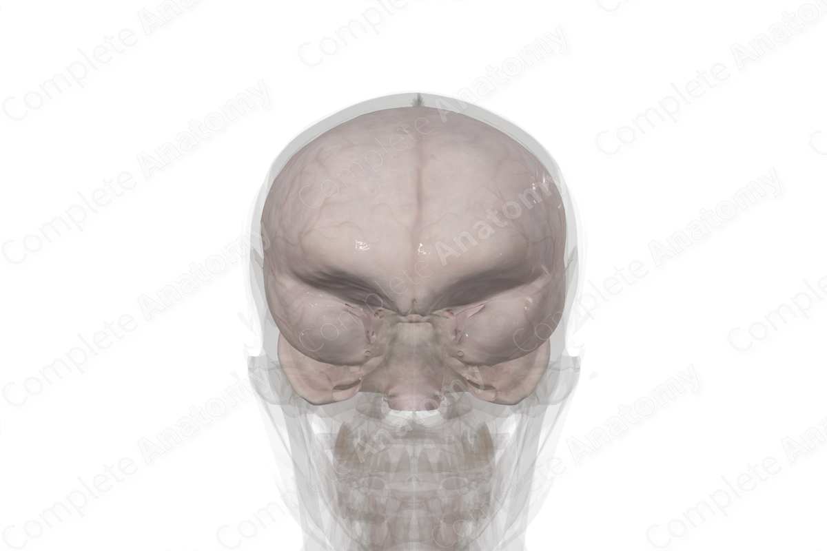
The cranial dura is the most superficial of the three meningeal layers and covers the entire brain down to the foramen magnum, where it is continuous with the spinal dura mater. The cranial dura mater is a tough, fibrous, two-layer structure with the outer or periosteal dura forming the periosteum of the cranial bones and the inner of meningeal dura laying against the underlying arachnoid mater. For the most part these two dural layers are adhered, but they can be visualized at some dural sinuses where the venous cavity separates the periosteal and meningeal dural layers. The dura is pierced by bridging veins, which carry blood from the brain; emissary veins, which bring blood from the overlying bone; and arachnoid granulations, which bring cerebrospinal fluid to venous sinuses.
Related parts of the anatomy
Anatomical Relations
The anterior, middle, and posterior meningeal arteries carry blood to the dura and overlying bone. The trigeminal and vagus nerves provide innervation to the vast bulk of the dura, with first to third cervical nerves innervating the dura mater near the foramen magnum.
List of Clinical Correlates
- Epidural hematoma
- Subdural hematoma
- Headache
Learn more about this topic from other Elsevier products
Dura Mater

A cephalocoele is a defect in the skull and dura with extra-cranial extension of intra-cranial structures.


