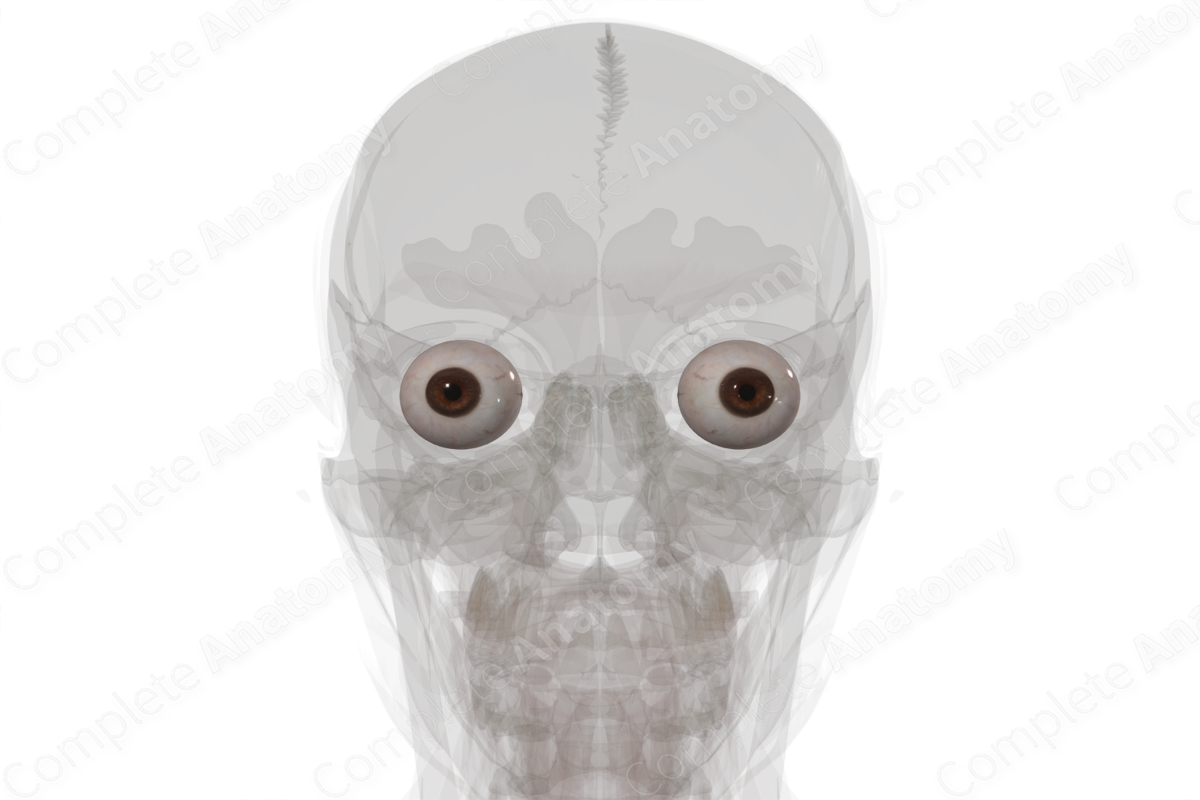
Structure
The eyeball is situated inside the anterior part of the orbit. It comprises of a multitude of structures, which are organized into three distinct layers.
- The outer fibrous layer comprises of cornea anteriorly and the sclera posteriorly.
- The middle vascular layer of the eyeball consists of the choroid, ciliary body, and iris.
- The inner layer is called the retina.


Key Features & Anatomical Relations
The cornea refers to the anterior one-sixth of the eyeball which is transparent in order to allow for the light to enter. On the contrary, the sclera forms the posterior five-sixths of the eyeball and is completely opaque. It is pierced by numerous vessels and nerve (including the optic nerve) and serves as the site of attachment of various muscles.
The choroid is a pigmented vascular layer which is attached to the retina internally and sclera externally. The ciliary muscle and ciliary processes protrude from the anterior border of the choroid and span in the form a complete ring around the eyeball. The muscle contributes to accommodation of the lens for near/far vision, while the ciliary processes produce aqueous humor.
Completing the vascular layer anteriorly is the iris. It projects outwards from the ciliary body as a circular structure and forms the colored part of the eye with a central pupillary opening inside it. Smooth muscle fibers in the iris control the size of the pupil.
The retina consists of an optic part anteriorly (concerned with vision) and a nonvisual part posteriorly, which underlines the ciliary body and the iris. Histologically, the optic part comprises of ten layers. The outermost is the pigmented layer, which is firmly attached to the choroid, while the inner neural layers contain nerve cells and fibers. The light sensitive receptors in the retina include cones (for light vision) and rods (for night vision). However, the area where the optic nerve exits the retina posteriorly, is devoid of photoreceptors and is called the optic disc. Lateral to the disc, is the site of maximum visual acuity, called the fovea. It contains a very high density of cones within it.
Learn more about this topic from other Elsevier products

Anatomy and physiology of the eye: Video, Causes, & Meaning

Anatomy and physiology of the eye: Symptoms, Causes, Videos & Quizzes | Learn Fast for Better Retention!
