Structure
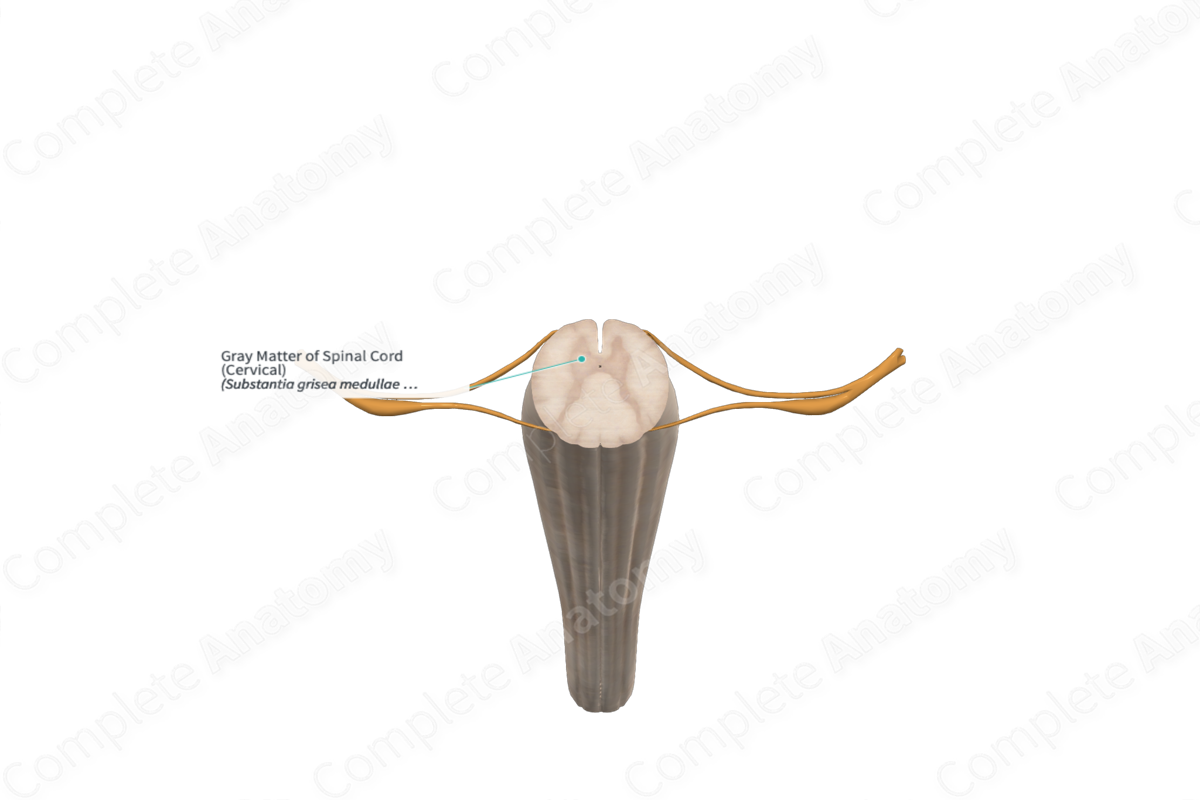
The spinal cord is composed of two types or regions, the inner gray matter and the surrounding white matter. Gray matter is rich in neuronal cell bodies and this results in a darker visual appearance in unstained tissue. When viewed in cross section, gray matter often appears to be shaped like a butterfly or an H.
Gray matter is often divided into different regions that are best seen when looking at cross sections. The dorsal horns run from the dorsolateral surface of the spinal cord and are the sites of sensory input to interneurons. This horn can be divided into different lamina, each of which are associated with different modalities and inputs.
The lateral horns, which are sometimes difficult to see, project almost directly laterally and are the site at which autonomic neuronal cell bodies are located.
The ventral horns, which form the thickest part of the gray matter and are located ventrally, are the site at which primary motor neurons are located. Motor and autonomic axons project out of the ventral horns in a ventrolateral direction.


Related parts of the anatomy
Key Features/Anatomical Relations
The gray matter forms an H or butterfly shape when seen in cross section. At the center lies the central canal, the evolutionary equivalent of the brain’s ventricles. This canal can carry cerebrospinal fluid through teen years, after which it is generally obliterated.
On both right and left sides, the gray matter has a dorsal horn corresponding to sensation, a lateral horn corresponding to autonomics, and a ventral horn corresponding to motor control. The dorsal horn can be subdivided into six different laminae, termed Rexed laminae. More commonly, three regions of the dorsal horn are discussed: the substantia gelatinosa, the nucleus proprius, and the dorsal nucleus.
The lateral horn has two Rexed laminae, but the most relevant feature is the intermediolateral nucleus.
The ventral horn also has two Rexed laminae and indistinct clusters of primary motor neuron cell bodies.
Learn more about this topic from other Elsevier products
Central Gray Matter

The spinal cord consists of central gray matter surrounded by columns of white matter.
