Structure
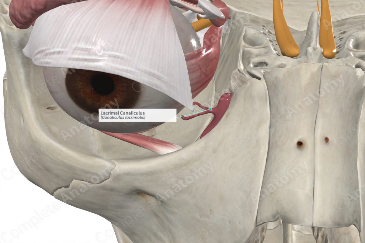
Each upper and lower eyelid contains a superior and inferior lacrimal canaliculus just adjacent to the medial angle of the eye. Tears collect at the medial angle of the eye and pass through a punctum to enter either the superior or inferior lacrimal canaliculus. Each canaliculus is initially vertical as it travels medially from its punctum. It then widens forming an ampulla and passes towards the lacrimal sac. The superior and inferior canaliculi unite forming a common lacrimal canaliculus, which drains into the lacrimal sac.


Related parts of the anatomy
Function
The lacrimal canaliculi are responsible for draining tears from the medial angle of the eye. Each tear drop consists of about 7μl of fluid. The tear is distributed over the cornea (1-2μl) and along the upper and lower margins of the eyelids (Standring, 2016). Some of the tear fluid is lost to evaporation or absorption into the conjunctiva, however, the majority is drained via the nasolacrimal drainage system. Tears collect at the medial angle of the eye (medial canthus) and two puncta in the region drain it into the superior and inferior lacrimal canaliculi. The canaliculi transport the tears to the lacrimal sac, and then onto the nasolacrimal duct which directs the tears to the inferior meatus of the nasal cavity.
References
Standring, S. (2016) Gray's Anatomy: The Anatomical Basis of Clinical Practice. Gray's Anatomy Series: Elsevier Limited.
