Quick Facts
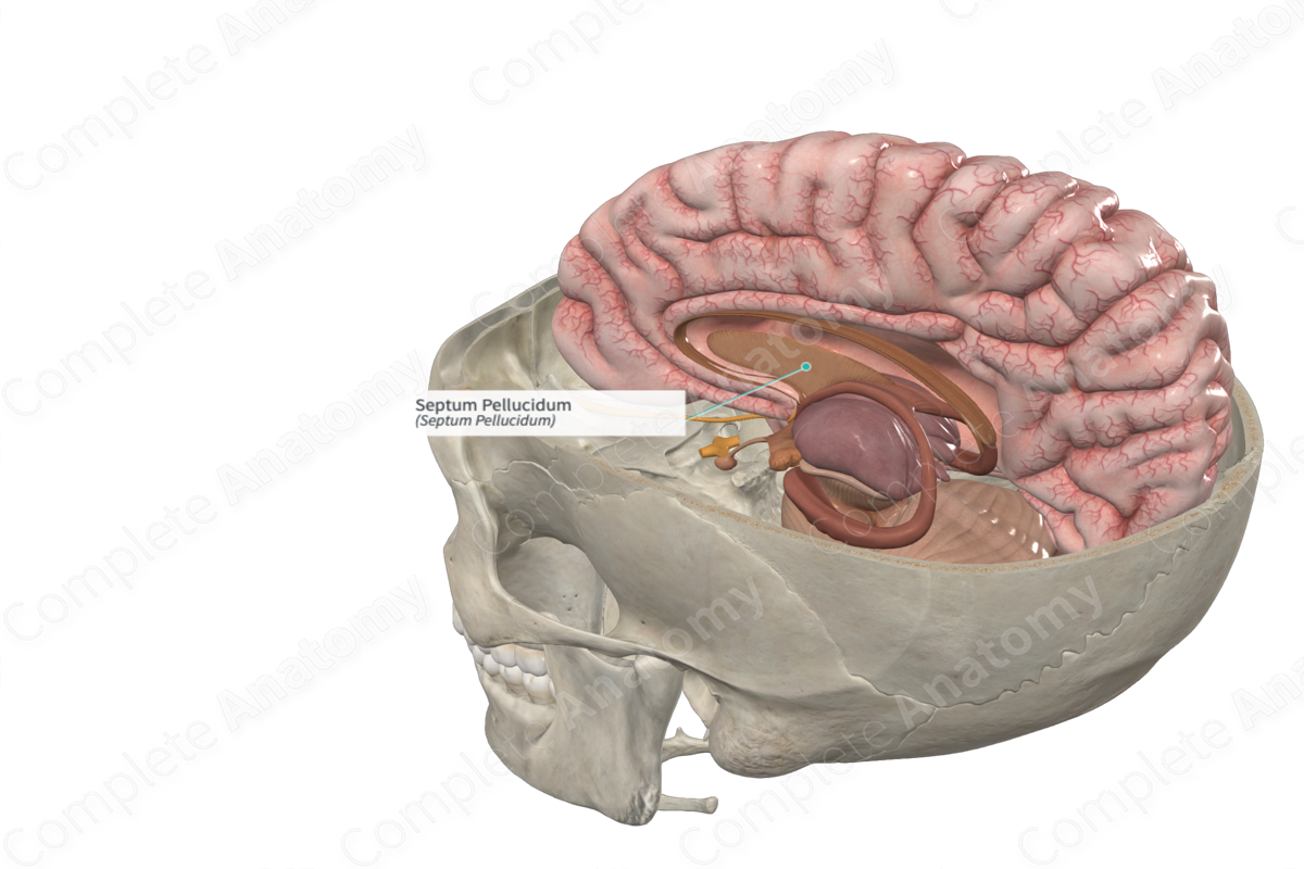
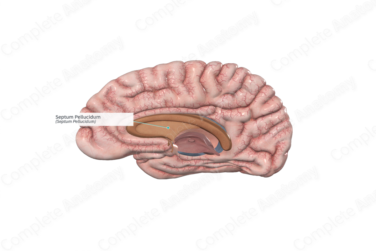
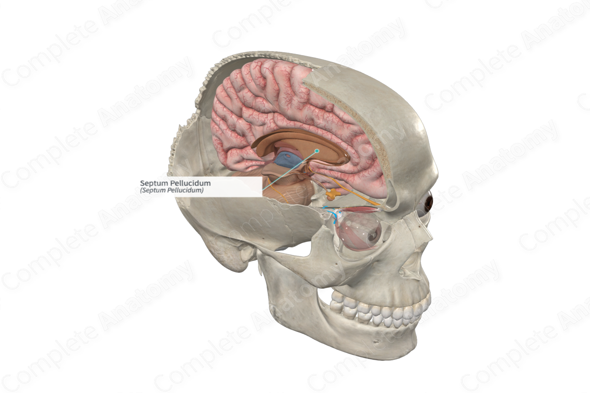
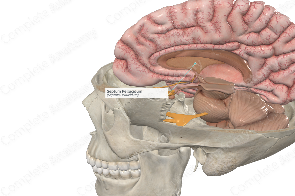
The septum pellucidum (aka septum lucidum) is a thin, triangular-shaped, membranous structure located between the two cerebral hemispheres. It separates the anterior horns and bodies of the lateral ventricles. The superior and anterior aspects of the septum pellucidum connect to the corpus callosum and the inferior and posterior aspects attach to the fornix.
The septum pellucidum consists of a right and left laminae, called the laminae septa pellucidi. During fetal development, the laminae are separated by a cavity called the cavum septa pellucidi (cavum meaning cavity or hollow). The cavity normally disappears during early infancy resulting in the fused septum pellucidum.
Absence of the septum pellucidum is a rare disorder and usually occurs with other neurological developmental abnormalities such as agenesis/dysgenesis of the corpus callosum. Absence of the septum pellucidum is commonly associated with vision impairment or blindness, hormone deficiencies resulting in short stature, coordination problems and learning disabilities.
Learn more about this topic from other Elsevier products


