
Structure/Morphology
The central nervous system develops as a tube in early embryonic life. While the brain undergoes dramatic enlargements, the spinal cord retains is tubular structure throughout life. The spinal cord is essentially a tube which runs from the base of the brainstem to the lumbar region. It is approximately 40-45 cm long. The spinal cord is divided up into 31 segments, each corresponding to a vertebral level and representing a spinal cord or neural level. There are 8 cervical, 12 thoracic, 5 lumbar, 5 sacral, and 1 coccygeal levels. Each level has a bilateral spinal nerve which gives rise to the peripheral nervous system.


Related parts of the anatomy
Key Features/Anatomical Relations
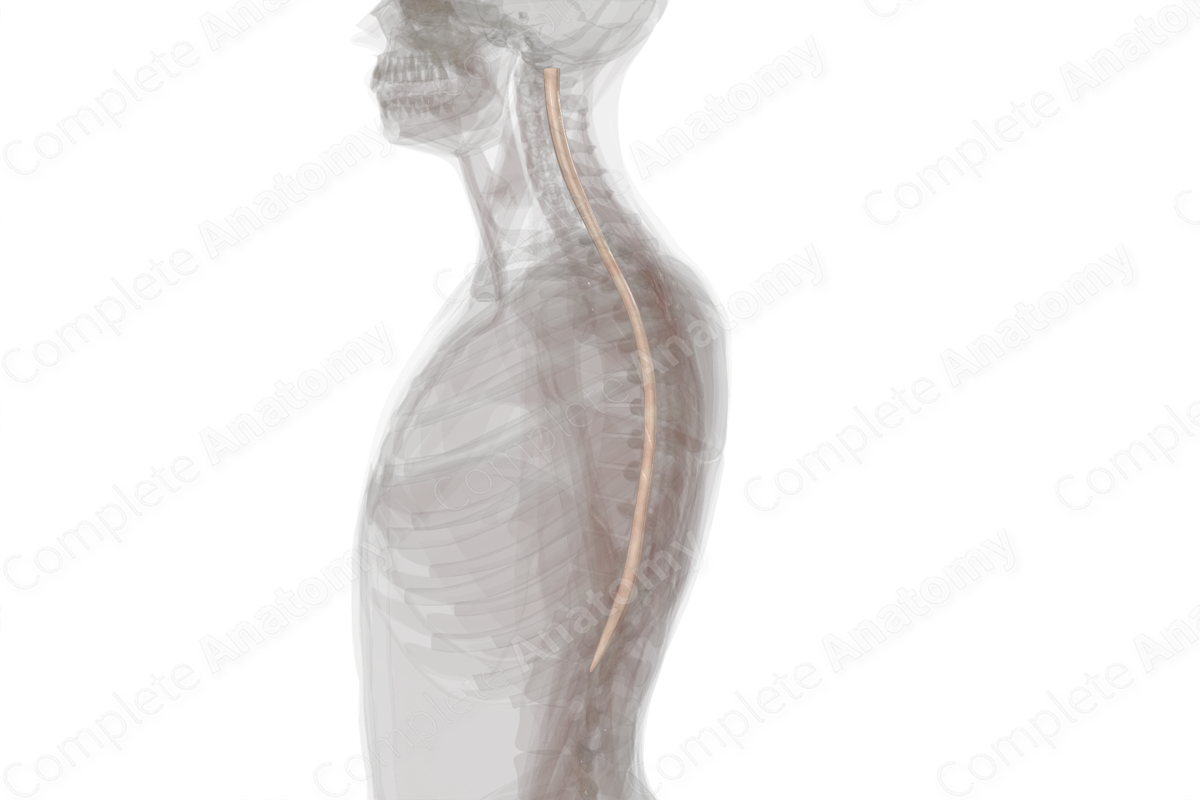
The spinal cord originates at the base of the brainstem, generally at the level of the foramen magnum. It extends inferiorly toward the pelvic region, always surrounded by the vertebral column and wrapped in the same three meningeal layers found in the brain: the inner pia, the middle arachnoid mater, and the outer dura mater. It is divided into different levels along the anterior/posterior axis corresponding to vertebral levels and anatomic targets: the cervical spinal cord serving the neck and upper limbs, the thoracic levels serving the trunk, and the lumbar and sacral levels serving the lower limbs. The cervical and lumbosacral levels are enlarged relative to the thoracic levels. Because the vertebral column grows more than the spinal cord, the spinal cord ends at a point called the conus medullaris, generally found between vertebral levels L1 and L2. Spinal nerve roots take on an increasingly downward course such that at cervical levels the nerve roots extend more laterally, while at lumbar levels the roots emerge from the spinal cord and run inferiorly.
When looking in cross section, the key features are gray matter and white matter. The gray matter holds the cell bodies and is found deep to the white matter. It takes on a butterfly appearance and can be divided into dorsal, lateral, and ventral horns. Surrounding the gray matter is the white matter. White matter is composed largely of the myelinated axons which convey sensory and motor information to and from the brain. Running down the center of the spinal cord is the central canal, a vestige from the development of the neural tube which is generally not patent in adults but can carry cerebrospinal fluid in earlier years. The central canal is continuous with the fourth ventricle, originating at the apex rostrally and ending in the conus medullaris caudally. Within the spinal cord, it is found centrally within the gray commissure. It is surrounded by gray matter that is classified as Rexed lamina X. The terminal expansion of the central canal is called the ventriculus terminalis of Krause.
Blood is supplied by the anterior spinal artery which runs in a groove in the ventral midline of the spinal cord, and the posterior spinal arteries which run on both the left and right sides of the dorsal midline.
List of Clinical Correlates
- Paralysis
- Spina bifida
- Lumbar puncture
Learn more about this topic from other Elsevier products

