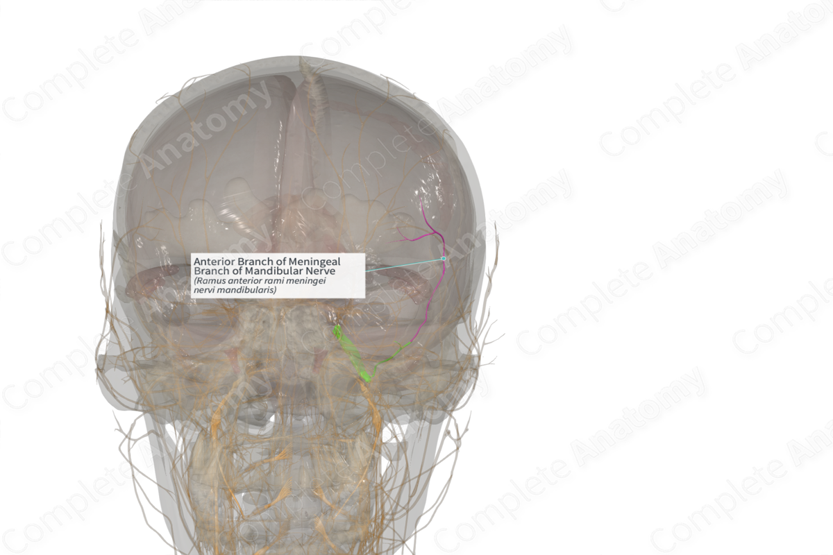
Anterior Branch of Meningeal Branch of Mandibular Nerve (Right)
Ramus anterior rami meningei nervi mandibularis
Read moreQuick Facts
Origin: Meningeal branch of trigeminal nerve.
Course: Roughly follows the middle meningeal artery branches that distribute to the dura mater of the middle cranial fossa.
Branches: None.
Supply: Conveys general sense fibers from the dura of the middle cranial fossa.

Related parts of the anatomy
Origin
The anterior branch of the meningeal branch of the trigeminal nerve is one of two branches. It originates intracranially after the meningeal branch of the trigeminal nerve passes through the foramen spinosum and bifurcates. Its sensory fibers have cell bodies located in the trigeminal ganglion.
Course
The anterior branch of the meningeal branch of the trigeminal nerve follows the branches of the middle meningeal artery to be distributed to dura of the middle cranial fossa.
Branches
There are no named branches, however, the anterior branch of the meningeal branch of the trigeminal nerve communicates with fibers from the meningeal branch of the maxillary nerve, which also innervates dura of the middle cranial fossa.
Supplied Structures
The anterior branch of the meningeal branch of the trigeminal nerve is a sensory nerve. It conveys general sense fibers from the dura of the middle cranial fossa.
Learn more about this topic from other Elsevier products
Mandibular Nerve

The marginal mandibular nerve is a branch of the inferior cervicofacial branch of the facial nerve that innervates the depressors of the lower lip, including the depressor anguli oris, depressor labii inferioris, and occasionally the pars labialis platysma.
