Quick Facts
Origin: First lumbar nerve (L1).
Course: Contributes to the lumbar plexus situated inside the psoas major muscle.
Branches: Iliohypogastric, ilioinguinal, and genitofemoral nerves.
Supply: Motor innervation to anterior abdominal wall musculature (external abdominal oblique, internal abdominal oblique, and transversus abdominis), cremaster muscle, and posterior abdominal wall musculature (psoas major, psoas minor, and quadratus lumborum). Sensory innervation to skin in the posterolateral gluteal region, pubic area, mons pubis, labium majus, upper medial, and anterior parts of thigh.


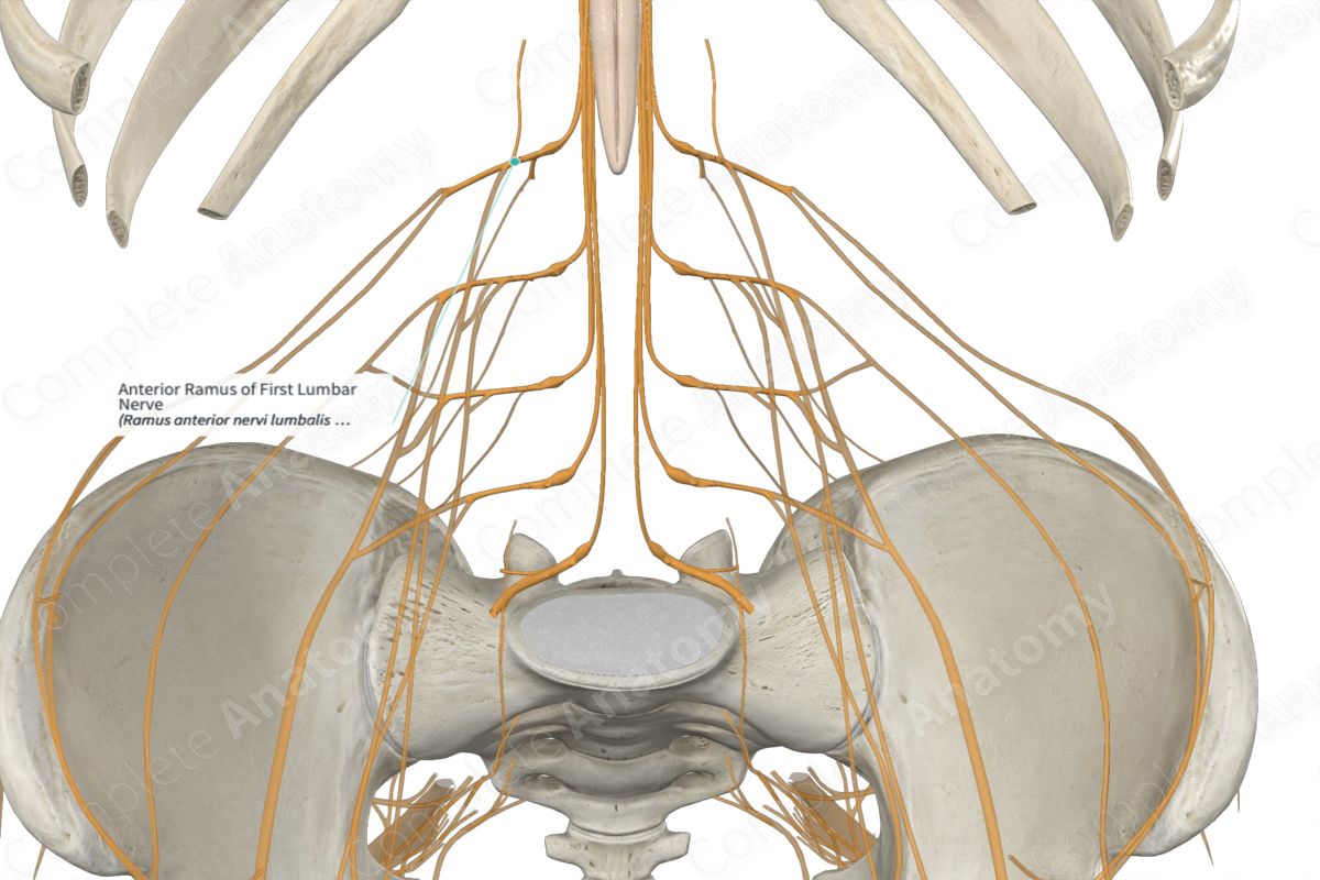
Origin
The anterior (ventral) ramus of first lumbar nerve originates as one of two branches of the first lumbar nerve. The other branch is the posterior (dorsal) ramus of the same L1 nerve.
Course
The bifurcation of the first lumbar nerve into anterior and posterior rami occurs immediately after it exits the intervertebral foramen. The anterior ramus contributes to the formation of the lumbar plexus, which also receives nerve fibers from the anterior rami of the second, third, and fourth lumbar, and subcostal nerves. The lumbar plexus is situated inside the substance of the psoas major muscle, anterior to its attachment to the transverse processes of the lumbar vertebrae.
Branches
The anterior ramus of first lumbar nerve is a mixed nerve which contains both somatic efferent (motor) and afferent (sensory) neurons.
The somatic efferent neurons emerge from the anterior gray horn of the L1 segment of the spinal cord. These are lower motor neurons which exit the spinal cord through the anterolateral sulcus as they travel inside the anterior motor rootlets and root of L1 spinal segment. They subsequently travel through the first lumbar nerve to enter the anterior ramus, before reaching the lumbar plexus. These efferent neurons travel through the branches of the lumbar plexus. These branches include the iliohypogastric, ilioinguinal, and genitofemoral nerves, which supply motor innervation to various structures.
Somatic afferent neurons travel through the lateral and anterior sensory cutaneous branches of the iliohypogastric and ilioinguinal nerves, and the genital and femoral branches of the genitofemoral nerve to enter the lumbar plexus. From here these neurons travel through the anterior ramus of the first lumbar nerve to enter the sensory root and rootlets of the first lumbar nerve. The cell bodies of these sensory neurons are located inside the spinal ganglion of the first lumbar nerve. The axons then travel through the posterolateral sulcus to enter the posterior sensory horn of the first lumbar spinal cord segment.
The anterior ramus of first lumbar nerve is also connected to the sympathetic trunk through the white and gray communicating branches (gray rami communicantes). These serve as conduits for the preganglionic and postganglionic sympathetic neurons, respectively.
Supplied Structures & Function
The anterior ramus of first lumbar nerve supplies motor innervation to the abdominal musculature (external abdominal oblique, internal abdominal oblique, and transversus abdominis muscles) through the somatic efferent neurons running inside the iliohypogastric and ilioinguinal nerves. They also provide motor innervation to the cremaster muscle through the genital branch of the genitofemoral nerve.
The somatic afferent neurons inside the iliohypogastric nerve conduct general sensory cutaneous information from the skin in the posterolateral gluteal and pubic regions. The somatic afferent neurons from the ilioinguinal nerve transmit general cutaneous sensory information from the upper medial thigh, mons pubis, and labium majus. While the somatic afferent neurons from the genitofemoral nerve bring general sensory cutaneous information from the skin of the upper anterior thigh and scrotum (males), mons pubis, or labium majus (females).
Some somatic afferents also supply sensory innervation to the anterior abdominal musculature through the iliohypogastric and ilioinguinal nerves (external abdominal oblique, internal abdominal oblique, and transversus abdominis muscles) and the posterior abdominal wall musculature (psoas major, psoas minor, and quadratus lumborum muscles) directly through the anterior ramus (Snell, 2011).
List of Clinical Correlates
—Lumbar sympathectomy
References
Snell, R. S. (2011) Clinical Anatomy by Regions. Lippincott Williams & Wilkins.
Learn more about this topic from other Elsevier products

