Quick Facts
Origin: Fourth lumbar nerve.
Course: Contributes to the lumbar plexus situated inside the psoas major muscle.
Branches: Femoral, obturator, superior gluteal, tibial, and common and deep fibular nerves.
Supply: Motor innervation to the posterior abdominal wall musculature (quadratus lumborum muscle), muscles of the anterior compartment of the thigh (sartorius, rectus femoris, vastus medialis, vastus intermedius, and vastus lateralis muscles) and adductor muscles (adductor longus, adductor magnus, gracilis, and obturator externus muscles). In addition, muscles in the gluteal region (gluteus medius, gluteus minimus, and tensor fasciae latae muscles) and leg region (popliteus, tibialis posterior, and tibialis anterior muscles) also receive motor innervation from the somatic efferent neurons of the anterior ramus of L4. Sensory innervation to the skin of the mons pubis, labium majus, anterior, medial, and lateral thigh regions, down to the level of the knee joint, and skin on the medial side of the leg, ankle, and foot. Moreover, articular branches innervate the hip and knee joints.


Origin
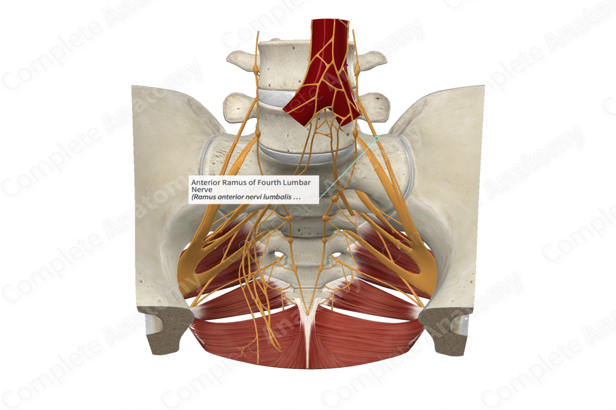
The anterior (ventral) ramus of fourth lumbar nerve originates as one of two branches of the fourth lumbar nerve. The other branch is the posterior (dorsal) ramus of the same L4 nerve.
Course
The bifurcation of the fourth lumbar nerve into anterior and posterior rami takes place immediately after its exit from the intervertebral foramen.
The anterior ramus of the fourth lumbar nerve contributes to the formation of the lumbar plexus, which also receives the nerve fibers from the anterior rami of the first, second, and third lumbar, and subcostal nerves.
Part of the anterior ramus of the fourth lumbar nerve combines with the anterior ramus of the fifth lumbar nerve to form the lumbosacral trunk, which courses vertically downwards into the pelvic cavity from the abdomen, by passing immediately anterior to the sacroiliac joint. The trunk contributes to the sacral plexus.
Branches
The anterior ramus of the fourth lumbar nerve is a mixed nerve which contains both somatic efferent (motor) and afferent (sensory) neurons.
The somatic efferent neurons emerge from the anterior motor rootlets and root of L4 spinal segment. They subsequently travel through the fourth lumbar nerve to enter the anterior ramus, before reaching the lumbar plexus. These efferent neurons travel through the muscular branches of the femoral and obturator nerves, to supply motor innervation to various muscles.
Remaining somatic efferent neurons in the anterior ramus of the fourth lumbar nerve pass through the lumbosacral trunk to enter the sacral plexus, and subsequently travel through branches of the plexus. For instance, efferent neurons enter the gluteal region, via the superior gluteal nerve, and supply the back of the knee joint and the leg, via the tibial, and common and deep fibular branches of the sciatic nerve.
Somatic afferent neurons travel through the cutaneous branches of the obturator and femoral nerves (medial, intermediate, and saphenous cutaneous nerves), to enter the lumbar plexus. From the here onwards these neurons travel through the anterior ramus of the fourth lumbar nerve to enter the sensory root and rootlets. The cell bodies of these neurons are located inside the spinal ganglion of the fourth lumbar nerve. The axons then travel through the posterolateral sulcus to enter the posterior sensory horn of the L4 spinal cord segment.
The anterior ramus of fourth lumbar nerve is also connected to the sympathetic trunk through the gray communicating branch (gray ramus communicans), which serves as a conduit for the postganglionic sympathetic neurons (Moore, Dalley and Agur, 2013).
Supplied Structures & Function
The anterior ramus of fourth lumbar nerve supplies motor innervation to the posterior abdominal wall musculature (quadratus lumborum) through the somatic efferent neurons coming directly from the ramus. In addition, these efferent neurons travel through the muscular branches of femoral nerve to innervates muscles of the anterior compartment of the thigh (sartorius, rectus femoris, vastus medialis, vastus intermedius, and vastus lateralis muscles). Moreover, efferent neurons from the anterior ramus the fourth lumbar nerve travel through muscular branches of obturator nerve to innervate muscles, including adductor longus, adductor magnus, gracilis, and obturator externus muscles.
Those efferent neurons that travel through the lumbosacral trunk, to enter the sacral plexus, supply somatic motor innervation to the gluteus medius, gluteus minimus, and tensor fasciae latae muscles via the superior gluteal nerve; the popliteus and tibialis posterior muscles via the tibial division of the sciatic nerve; the tibialis anterior muscle via the common fibular division of the sciatic nerve.
The somatic afferent neurons from the medial, intermediate, and saphenous cutaneous branches of the femoral nerve transmit general sensations from the anterior and medial parts of thigh, and skin on the medial side of the leg, ankle, and foot. Some neurons exit from the obturator and the femoral nerves to innervate the hip and the knee joints (Moore, Dalley and Agur, 2013).
List of Clinical Correlates
—Laminectomy
—Lumbosacral plexopathy (Planner, Donaghy and Moore, 2006)
—Back pain (Moore, Dalley and Agur, 2013)
References
Moore, K. L., Dalley, A. F. and Agur, A. M. R. (2013) Clinically Oriented Anatomy. Clinically Oriented Anatomy 7th edn.: Wolters Kluwer Health/Lippincott Williams & Wilkins.
Planner, A. C., Donaghy, M. and Moore, N. R. (2006) 'Causes of lumbosacral plexopathy', Clin Radiol, 61(12), pp. 987-95.
Learn more about this topic from other Elsevier products

