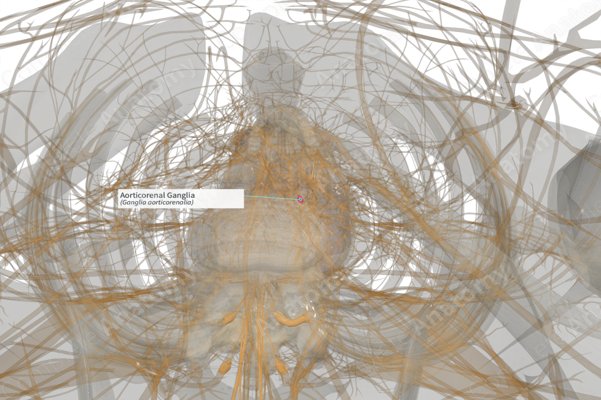
Description
The aorticorenal ganglion is one of two paired prevertebral abdominal sympathetic ganglia. The ganglia are typically found at the confluence of the renal arteries and abdominal aorta. Often, the aorticorenal ganglia are partly fused to the celiac ganglion and can be considered its inferior extension. Primarily, the aorticorenal ganglion receives input from sympathetic preganglionic neurons located in the intermediolateral cell column of twelfth thoracic nerve (T12), by way of the least thoracic splanchnic nerve.
However, some contribution may arise from eleventh thoracic or first lumbar nerves (T11 or L1) and may travel through the lesser thoracic splanchnic nerve or first lumbar splanchnic nerve, respectively.
The ganglia contribute postganglionic sympathetic nerve fibers to a number of nearby periarterial plexuses, including the celiac, superior mesenteric, renal, aortic, and gonadal plexuses.
The broad and diverse distribution of postganglionic sympathetic axons from the aorticorenal ganglion ensures that it has a multitude of targets and effects. These include the kidneys, suprarenal glands, superior portion of the ureter, and gonads (Norvell, 1968).
Parasympathetic effector and visceral sensory afferent fibers pass through the aorticorenal ganglion on their way to or from the kidneys, ureter, and gonads. However, they do not synapse in the aorticorenal ganglion.
References
Norvell, J. E. (1968) 'The aorticorenal ganglion and its role in renal innervation', J Comp Neurol, 133(1), pp. 101-11.


