Quick Facts
Sympathetic Contribution: Cardiac plexuses.
Parasympathetic Contribution: Cardiac plexuses.
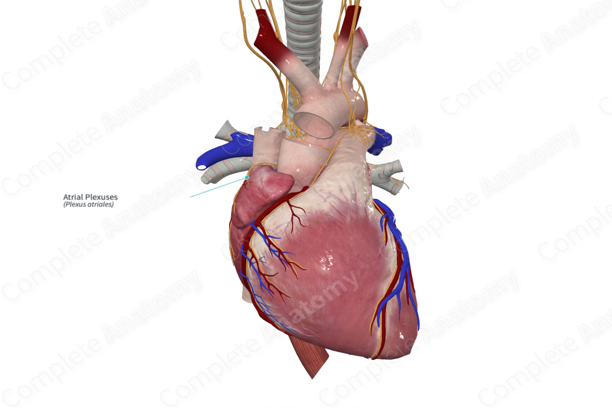
Course: Lay clustered on the anterior, superior, or posterior surfaces of the atria.
Sympathetic Supply: Increases heart rate, cardiac impulse conduction, and force of myocardial contraction, dilates coronary arteries.
Parasympathetic Supply: Decrease heart rate and force of myocardial contraction.
Contributing Nerves
The atrial plexuses represent a continuation of the lateral aspects of both the superficial and deep cardiac plexuses.
Course
The atrial plexuses are found on the surface of the left and right atria, and typically, there are five plexuses (Armour et al., 1997, Qin et al., 2017).
Branches
The atrial plexuses have no named branches. Fibers leave the plexuses to innervate cardiac tissue in the atria.
Supplied Structures & Function
The atrial plexuses provide both sympathetic and parasympathetic innervation to the heart, particularly the atria. Both types of nerve fibers pass from the plexus into the cardiac tissue, primarily in the atrial regions.
The sympathetic fibers lead to dilation of coronary arteries and increased rate and force of cardiac myofiber contraction. Parasympathetic fibers do the opposite, constricting coronary arteries and reducing heart rate.
Visceral sensory fibers traveling through the atrial plexus convey information relating to pain, blood pressure, and blood chemistry back to the central nervous system.
List of Clinical Correlates
—Referred pain
References
Armour, J. A., Murphy, D. A., Yuan, B. X., Macdonald, S. and Hopkins, D. A. (1997) 'Gross and microscopic anatomy of the human intrinsic cardiac nervous system', Anat Rec, 247(2), pp. 289-98.
Qin, M., Zhang, Y., Liu, X., Jiang, W. F., Wu, S. H. and Po, S. (2017) 'Atrial Ganglionated Plexus Modification: A Novel Approach to Treat Symptomatic Sinus Bradycardia', JACC Clin Electrophysiol, 3(9), pp. 950-959.
