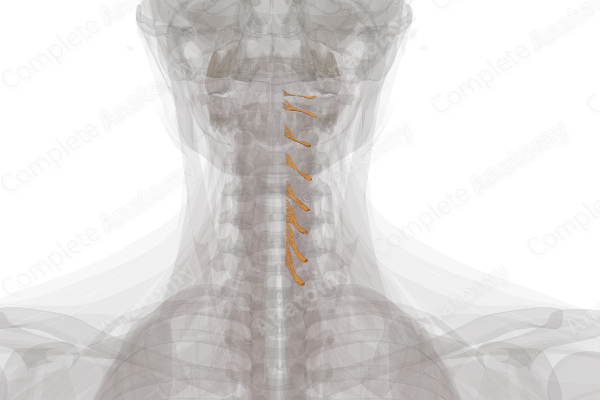
Cervical Spinal Roots & Ganglia (Left)
Radicis et ganglia spinalia cervicales
Read moreDescription
A spinal nerve emerges bilaterally at each cervical spinal cord level. There are eight pairs of cervical spinal nerves, each of which exit the intervertebral foramina above their corresponding cervical vertebra. However, since there are only seven cervical vertebrae, the eighth cervical spinal nerve exits the intervertebral foramen below the seventh cervical vertebra. From here below, each spinal nerve (12 thoracic, 5 lumbar, 5 sacral) emerges below its corresponding thoracic, lumbar, and sacral vertebrae.
Like other spinal nerves, cervical nerves are “mixed nerves” and contain both afferent (sensory) and efferent (motor) axons, which transmit information traveling towards or away from the central nervous system (CNS), respectively. Shortly after a cervical nerve emerges from the spinal cord it divides into posterior (dorsal) and anterior (ventral) rami. The posterior cervical rami innervate the muscles and skin over the back of the neck and scalp, while the anterior rami innervate skin covering the lateral and anterior portions of the neck and the angle of the mandible.
The cervical nerve roots represent the axons that enter or exit the spinal cord in the cervical region. All sensory (afferent) information enters the spinal cord via the posterior roots and all motor (efferent) information exits the spinal cord via the anterior roots. Each cervical nerve is formed by the union of one posterior and one anterior root at its respective spinal level.
The spinal ganglia are located at the proximal ends of the posterior roots, either inside or just exterior to the intervertebral foramina. Ganglia are swellings in which the cell bodies of afferent sensory neurons are situated.


Related parts of the anatomy
Learn more about this topic from other Elsevier products
Cervical Nerve Root

Technically the cervical nerve “root” consists of that portion of each nerve arising from the spinal cord and lying in the subdural space.
