Description
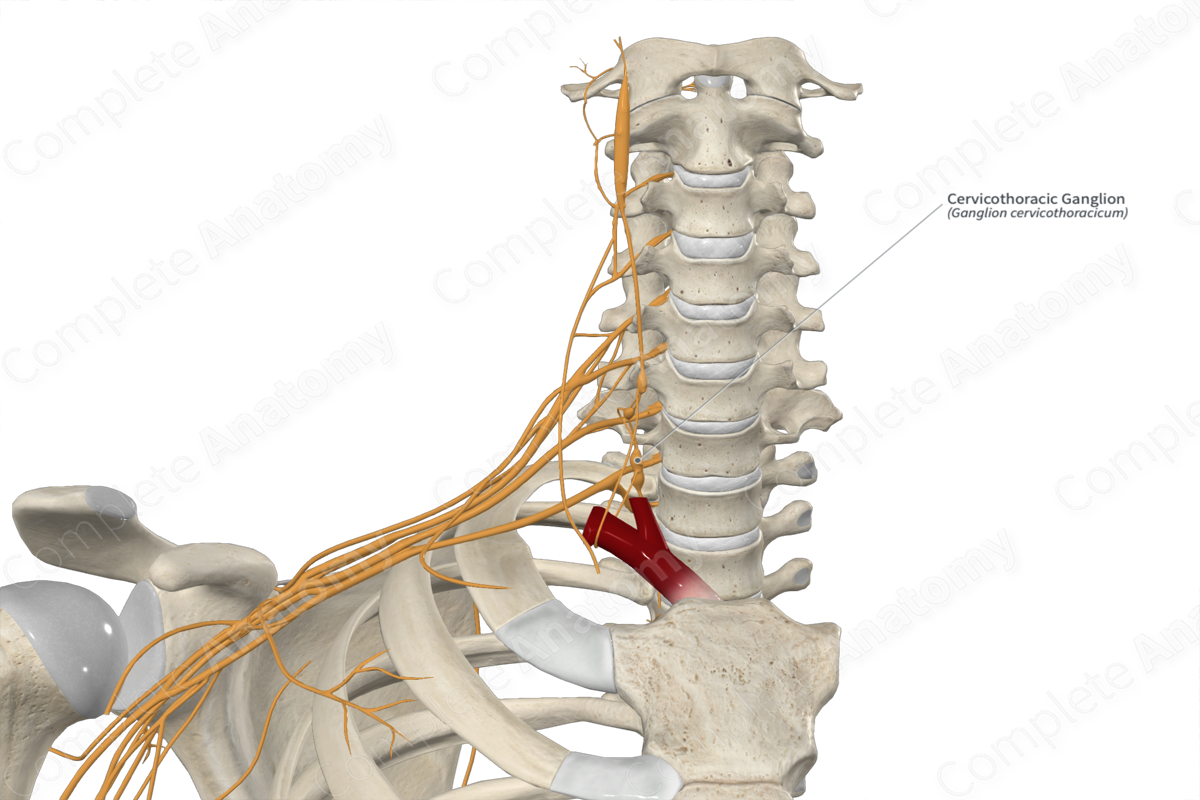
The cervicothoracic ganglion, also known as the stellate ganglion, is typically a fusion of the most inferior cervical ganglion and the most superior thoracic ganglion of the sympathetic chain. It is a bilateral paravertebral ganglion that contains postganglionic sympathetic neuronal cell bodies and transmits preganglionic sympathetic axons.
The cervicothoracic ganglion is located anterior to the neck of the first rib, posterior to the origin of the vertebral artery, and superior to the pleura. It lies medial to the scalene muscles and lateral to the longus colli muscle.
Fibers leaving the cervicothoracic ganglion travel along the vertebral and subclavian arteries, gray rami communicantes to the anterior rami of the sixth to eighth cervical nerves, and the inferior cervical cardiac nerve to the cardiac plexuses. Targets of the cervicothoracic ganglion include the head, neck, upper limbs, and thorax (Marcer et al., 2012).
List of Clinical Correlates
—Raynaud’s disease
—Hyperhydrosis
References
Marcer, N., Bergmann, M., Klie, A., Moor, B. and Djonov, V. (2012) 'An anatomical investigation of the cervicothoracic ganglion', Clin Anat, 25(4), pp. 444-51.
Learn more about this topic from other Elsevier products
Stellate Ganglion

The stellate ganglion, also known as the cervicothoracic ganglion, represents a fusion of the inferior cervical and first thoracic ganglia of the sympathetic trunk.


