Quick Facts
Sympathetic Contribution: The sympathetic neurons for the coccygeal, fifth and fourth sacral anterior rami arise from the lateral horns of the lumbar spinal cord segments.
Parasympathetic Contribution: The parasympathetic neurons, from the lateral horn of fourth sacral spinal segment, travel through the anterior ramus of fourth sacral nerve, before exiting through the pelvic splanchnic nerves (Note: Pelvic splanchnic nerves exit from the anterior rami of S2—S4).
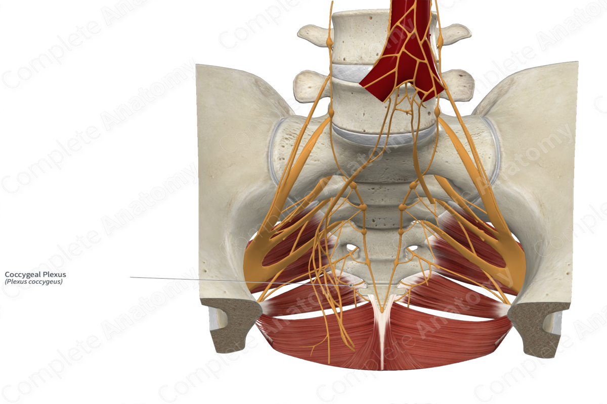
Course: The anterior rami of fifth sacral and coccygeal nerve penetrate the coccygeus muscle and join with the anterior ramus of fourth sacral nerve to form the coccygeal plexus.
Sympathetic Supply: Gray communicating branches connect the anterior rami of the coccygeal, and fifth and fourth sacral nerves with the sympathetic trunk and serve as conduits for the postganglionic sympathetic neurons.
Parasympathetic Supply: The preganglionic parasympathetic neurons in the pelvic splanchnic nerve synapse inside the inferior hypogastric plexus. They supply pelvic structures, including the urinary bladder, prostate, seminal vesicles, rectum, and uterus, penis, or clitoris.


Contributing Nerves
The coccygeal plexus is formed by the contributions from the following:
—a minor contribution from the anterior ramus of the fourth sacral nerve (S4);
—a major contribution from the anterior rami of fifth sacral nerve (S5), the coccygeal nerve (Co);
—a sympathetic contribution from gray rami.
The cell bodies are situated inside the ganglia of the sympathetic trunks. The cell bodies of the preganglionic sympathetic neurons are situated inside the lateral gray horn of the spinal cord segments.
Course
The anterior rami of fifth sacral and coccygeal nerve exit the sacral hiatus at a level caudal to the pelvic floor. They pass anteriorly and superiorly to penetrate the coccygeus muscle and enter the pelvic cavity. Here they join with the anterior ramus of fourth sacral nerve to form the coccygeal plexus. Some consider the plexus to form within the body of the coccygeus muscle (Woon and Stringer, 2014).
Branches
Small anococcygeal nerves originate from the coccygeus plexus. They penetrate the coccygeus muscle and the overlying sacrospinous and sacrotuberous ligaments. They pass superficially and innervate the skin in the anal triangle of the perineum (perianal skin) and coccygeal region.
Preganglionic parasympathetic neurons from the lateral horn of the fourth sacral spinal cord segment travel through the anterior ramus of fourth sacral and coccygeal plexus. These travel through the pelvic splanchnic nerve to synapse with the postganglionic parasympathetic neuronal cell body inside the inferior hypogastric plexus. These postganglionic neurons supply the pelvic viscera and external genitalia.
Supplied Structures
The coccygeal plexus provides sensory innervation to the perianal skin (over the anal triangle of the perineum) and the coccygeal region through the anococcygeal nerves.
It also provides parasympathetic motor innervation to the pelvic structures, such as the urinary bladder, prostate, seminal vesicles, rectum, uterus, and penis, or clitoris.
Sympathetic innervation to the pelvic region is distributed into the anterior rami through the gray communicating branches (gray rami communicantes containing postganglionic sympathetic fibers).
List of Clinical Correlates
—Coccydynia
References
Woon, J. T. and Stringer, M. D. (2014) 'Redefining the coccygeal plexus', Clin Anat, 27(2), pp. 254-60.
Learn more about this topic from other Elsevier products

