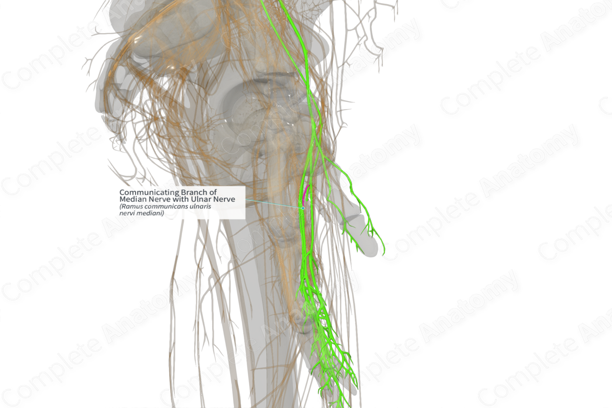
Communicating Branch of Median Nerve with Ulnar Nerve (Left)
Ramus communicans ulnaris nervi mediani
Read moreQuick Facts
Origin: Neuronal interconnections between the ulnar and median nerves.
Course: Within the axilla, cubital fossa, distal forearm, or hand.
Branches: None.
Supply: Median and ulnar nerves innervate the arm (flexor compartment) and hand regions. The neuronal interconnections between the median and ulnar nerves alter the normal neuronal distribution leading to difficulties in clinical interpretation.
Related parts of the anatomy
Origin
Anastomosis or neuronal interconnections between the ulnar and median nerves have been reported at various levels, for instance:
—in the axilla or upper arm;
—in the cubital fossa;
—distal forearm region;
—superficial palmar region, both proximally and distally (Roy et al., 2016).
Course
The first communication between the median nerve and the ulnar nerve has been observed at/immediately distal to the union of lateral and medial roots of the median nerve (Mat Taib et al., 2017).
The intercommunicating branch between the two nerves in the cubital fossa is called the Martin-Gruber anastomosis. It emerges as a neuronal communication from the main trunk of median nerve or the anterior interosseous nerve. It crosses superficial to the flexor digitorum superficiales or flexor digitorum profundus muscles, before it ends up by joining the ulnar nerve.
Another infrequent neuronal anastomosis can sometimes be observed between ulnar and median nerves in the distal forearm, also known as the Marinacci anastomosis.
The fourth type of neural connection is a palmar anastomosis between the recurrent branch of the median nerve and the deep branch of the ulnar nerve, leading to complete innervation of the thenar muscles by the ulnar nerve. It is also called the Riche-Cannieru anastomosis.
The fifth type of neuronal anastomosis is a communication in the palmar surface of the hand, between the common digital nerve arising from the median and ulnar nerves.
Branches
There are no named branches.
Supplied Structures
Broadly speaking, the median nerve is the predominant source of innervation for the flexor compartment of the forearm, with minor contributions from the ulnar nerve (medial parts of flexor digitorum profundus and flexor carpi ulnaris). The ulnar nerve serves as the predominant source of innervation for the musculature of the hand, with lesser contributions from the median nerve. The neuronal interconnections between the median and ulnar nerves alter the normal neuronal distribution leading to difficulties in clinical interpretation described below.
These anastomoses are often asymptomatic and remain undiscovered until nerve injuries to the ulnar or median nerves occur and lead to unusual motor or sensory deficits. Because they are clinically silent, their presence can cause difficulties in interpreting electrophysiological findings for the diagnosis of neuropathy and are at risk for iatrogenic damage during surgeries. Nerve entrapment disorders, such as carpal tunnel syndrome, has been known to be exacerbated or diminished in the setting of these neuronal anastomoses, leading to misdiagnosis and improper treatment.
References
Mat Taib, C. N., Hassan, S. N., Esa, N., Mohd Moklas, M. A. and San, A. A. (2017) 'Anatomical variations of median nerve formation, distribution and possible communication with other nerves in preserved human cadavers', Folia Morphol (Warsz), 76(1), pp. 38-43.
Roy, J., Henry, B. M., PĘkala, P. A., Vikse, J., Saganiak, K., Walocha, J. A. and Tomaszewski, K. A. (2016) 'Median and ulnar nerve anastomoses in the upper limb: A meta-analysis', Muscle Nerve, 54(1), pp. 36-47.


