Quick Facts
Sympathetic Contribution: Cervical and thoracic cardiac nerves of the sympathetic trunk.
Parasympathetic Contribution: Vagus and recurrent laryngeal nerves.
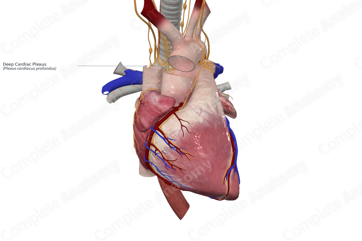
Course: Lies posterior to the aortic arch and anterior to the trachea, roughly at the level of the tracheal bifurcation.
Sympathetic Supply: Increases heart rate, cardiac impulse conduction, and force of myocardial contraction, dilates coronary arteries.
Parasympathetic Supply: Decreases heart rate and force of myocardial contraction.


Contributing Nerves
The deep cardiac plexus is formed by cardiac branches that extend from the vagus nerves and the sympathetic trunks.
The sympathetic component of the deep cardiac plexus comes from all the cardiac nerves except the left superior cervical cardiac nerve (Standring, 2016). These nerves include:
—right superior, middle, inferior cervical cardiac nerves;
—left middle and inferior cardiac cervical nerves;
—Left and right thoracic cardiac branches.
The preganglionic sympathetic cell bodies reside in the lateral horn of the T1 through T5 spinal cord segments. The postganglionic sympathetic cell bodies reside in the superior (right), middle, and inferior cervical ganglia, as well as the uppermost thoracic ganglion if distinct from the cervicothoracic ganglion.
The parasympathetic component of the deep cardiac plexus comes from all the cardiac branches of the vagus nerve except the inferior cervical cardiac branch of the left vagus nerve (Standring, 2016). These nerves include the:
—superior and inferior cervical cardiac branches of the right vagus nerve;
—inferior cervical cardiac branches of the left vagus nerve;
—thoracic cardiac branches of the vagus nerve;
—branches of the recurrent laryngeal nerves.
The preganglionic parasympathetic cell bodies reside in the dorsal nucleus of the vagus nerve, in the medulla oblongata. The postganglionic parasympathetic cell bodies reside in the various cardiac ganglia spread throughout the cardiac plexuses and cardiac tissue.
Course
The deep cardiac plexus is located posteriorly to the aorta and anterior to the trachea, roughly at the level of the tracheal bifurcation.
Branches
The deep cardiac plexus gives off the following branches:
—atrial plexuses;
—left coronary plexus;
—right anterior pulmonary plexus;
—right coronary plexus.
The deep cardiac plexus is also in communication with the superficial cardiac plexus through extensive mixing of fibers (Standring, 2016).
Supplied Structures & Function
The deep cardiac plexus provides both sympathetic and parasympathetic innervation to the heart, particularly the atria. Both types of nerve fibers pass from the plexus into the cardiac tissue, primarily in the atrial regions.
The sympathetic fibers lead to dilation of coronary arteries and increased rate and force of cardiac myofiber contraction. Parasympathetic fibers do the opposite, constricting coronary arteries and reducing heart rate.
Visceral sensory fibers traveling through the deep cardiac plexus convey information relating to pain, blood pressure, and blood chemistry back to the central nervous system.
List of Clinical Correlates
—Referred pain
References
Standring, S. (2016) Gray's Anatomy: The Anatomical Basis of Clinical Practice. Gray's Anatomy Series: Elsevier Limited.
Learn more about this topic from other Elsevier products
Plexus

Visceral plexuses are a network of nerve fiber and ganglia surrounding organs of the abdomen and pelvis region that convey sympathetic, parasympathetic, and visceral afferent input.
