Quick Facts
Origin: First thoracic nerve.
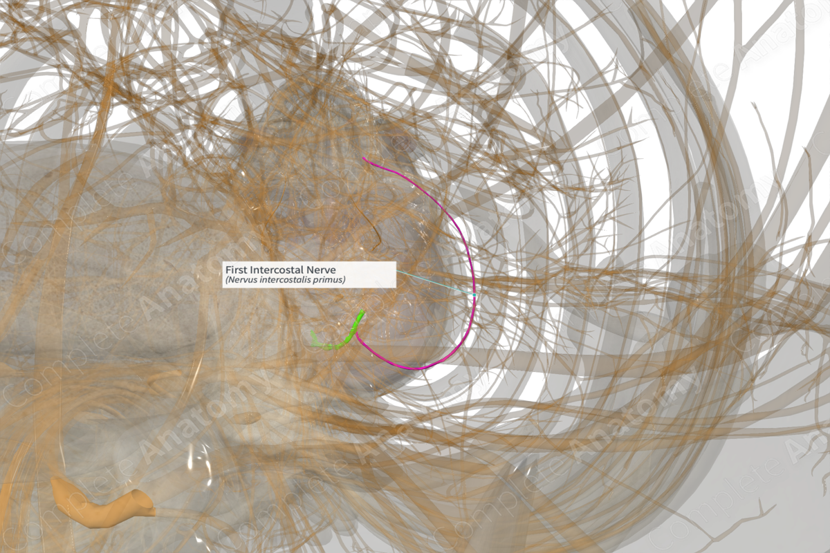
Course: Travels within the first intercostal space.
Branches: Collateral, lateral cutaneous, and anterior cutaneous branches.
Supply: The anterior ramus supplies motor innervation to the muscles of the first intercostal space and conveys sensory innervation from the skin overlaying the first intercostal space.


Related parts of the anatomy
Origin
The majority of the nerve fibers from the anterior ramus of the first thoracic nerve contributes to the brachial plexus as the inferior root of the inferior trunk. The remaining fibers continue as the first intercostal nerve.
Course
As the first intercostal nerve arises, it travels within the first intercostal space, close to the inferior border of the first rib and sits between the internal intercostal and the innermost intercostal muscles. It is also accompanied by an intercostal artery and vein, where the vein sits closest to the rib above, followed by the artery and nerve.
Branches
Near its origin, the first intercostal nerve gives off a collateral branch that descends in the intercostal space and courses along the inferior border of the space, in the same plane as the first intercostal nerve.
A lateral cutaneous branch arises near the mid-axillary line and pierces the chest wall anterior to the serratus anterior muscle.
The anterior cutaneous branch arises as the first intercostal nerve reaches the anterior portion of the intercostal space. The anterior cutaneous branch also pierces the chest wall.
Supplied Structures & Function
The first intercostal nerve supplies motor innervation to the intercostal muscles of the first intercostal space (external intercostal, internal intercostal, innermost intercostal), and sensory innervation to the skin overlying the first intercostal space.
Learn more about this topic from other Elsevier products

