Quick Facts
Origin: Preganglionic efferent fibers from thoracic and lumbar splanchnic nerves and preaortic ganglia send postsynaptic sympathetic fibers to the iliac plexus.
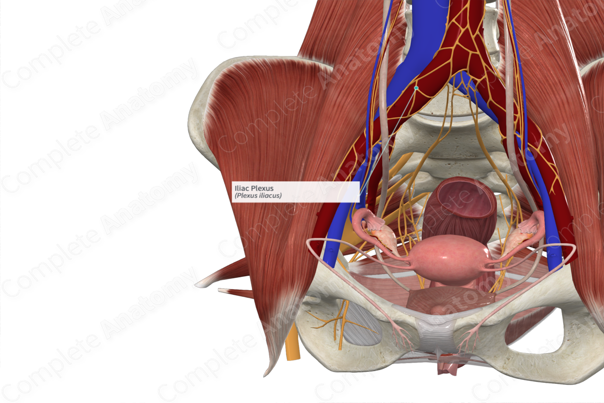
Course: Lies bilaterally on the left and right iliac arteries.
Branches: None.
Supply: Likely to innervate the vascular smooth muscle and tissues of the pelvis and lower limbs.
Origin
The iliac plexus is contiguous with the aortic plexus and the inferior hypogastric plexus. It likely receives fibers from thoracic and lumbar splanchnic nerves and preaortic ganglia. It is unclear if any parasympathetic or visceral sensory fibers contribute to the iliac plexus.
Course
The iliac plexus lies on the common iliac artery and the external and internal iliac arteries.
Branches
The iliac plexus does not give rise to any named branches.
Supplied Structures
The iliac plexus is thought to be largely a plexus of sympathetic fibers. Sympathetic efferent fibers are thought to innervate vascular smooth muscle and tissues of the lower limb and pelvis.
It is unclear if any parasympathetic or visceral sensory fibers contribute to the iliac plexus.
Learn more about this topic from other Elsevier products


