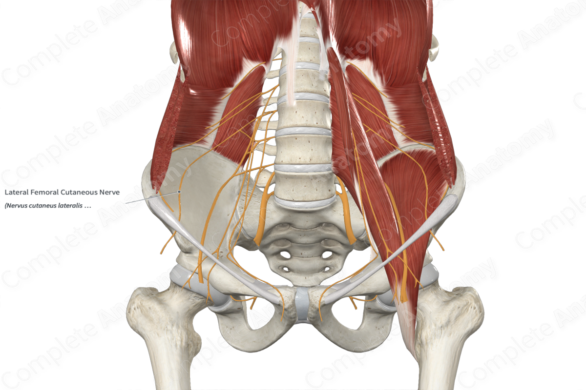
Quick Facts
Origin: Lumbar plexus (L2—L3).
Course: Emerges from the lateral border of the psoas major, traverses over the iliacus muscle before passing underneath the inguinal ligament medial to the anterior superior iliac spine.
Branches: None.
Supply: Sensory innervation for the skin of the anterior and posterior aspects of the lateral thigh, as far inferiorly as the knee.
Related parts of the anatomy
Origin
The lateral femoral cutaneous nerve is formed inside the psoas major muscle by the posterior divisions of the anterior rami of second and third lumbar nerves (L2—L3).
Course
Following its origin, the lateral femoral cutaneous nerve emerges from the lateral border of the psoas major muscle and courses obliquely downwards across the iliacus muscle. The nerve travels deep to iliacus fascia as it descends towards the anterior superior iliac spine (ASIS). It passes posterior to the inguinal ligament and enters the thigh just medial to the ASIS. It traverses over the sartorius muscle to lie on its anterior surface.
Branches
The lateral femoral cutaneous nerve has no named branches; however, its terminal ramifications extend in anterior and posterior directions to supply the anterior and posterior aspects of the lateral thigh, as far inferiorly as the knee. This is a pure sensory nerve and contains only somatic afferent (sensory) neurons.
The anterior ramifications contain mostly L3 neuronal fibers and are distributed along the anterior and lateral parts of the skin of the thigh. A few terminal neurofilaments also contribute to the prepatellar plexus (along with the infrapatellar branch of saphenous nerve) (Anloague and Huijbregts, 2009). The posterior ramifications contain mostly L2 neuronal fibers and are distributed along the lateral and posterior parts of the skin of the thigh.
Supplied Structures
The lateral femoral cutaneous nerve provides sensory innervation to the skin of the anterior and posterior aspects of the lateral thigh, as far inferiorly as the knee.
List of Clinical Correlates
—Meralgia paresthetica
References
Anloague PA, Huijbregts P. Anatomical variations of the lumbar plexus: a descriptive anatomy study with proposed clinical implications. J Man Manip Ther. 2009;17(4):e107-14.


