Quick Facts
Sympathetic Contribution: The first and second lumbar nerve cell bodies (L1-L2) descend in the sympathetic trunk and exit with second to fourth sacral nerves. (S2-S4) Some of these terminate on corresponding paravertebral ganglia, but most travel as sacral splanchnic nerves to synapse in prevertebral ganglia embedded within the inferior hypogastric plexus.
Parasympathetic Contribution: Pelvic splanchnic nerves (S1—S4).
Course: Both sympathetic and parasympathetic nerves pass through the inferior hypogastric plexus and are distributed to targets by contributing to the periarterial plexus surrounding the middle rectal artery.
Sympathetic Supply: Promotes continence by innervating smooth muscle of the rectal wall and internal anal sphincter.
Parasympathetic Supply: Promoting defecation by innervating smooth muscle of the rectal wall and internal anal sphincter.


Contributing Nerves
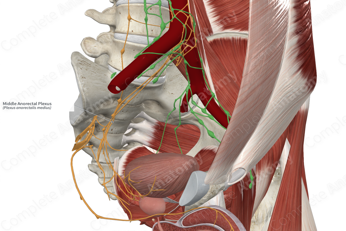
The middle anorectal plexus is formed by sacral (sympathetic) and pelvic (parasympathetic) splanchnic nerves. These axons pass to the inferior hypogastric plexus, where most of the sympathetic nerves synapse. The territory of autonomic nerves of the middle rectum overlaps with the superior rectal plexus. Therefore, the middle plexus may receive some input via these nerves.
Course
The nerves of the middle anorectal plexus pass to the rectum, either extraperitoneally, directly, or by contributing to the periarterial plexus surrounding the middle rectal artery.
Branches
There are no named branches of this dense and extensive plexus.
Supplied Structures
The autonomic fibers of the middle anorectal plexus supply smooth muscle of the rectum, particularly the middle portion. The smooth muscle includes the muscularis layer (longitudinal muscle) and the inferior anal sphincter.
Visceral sensory fibers from the rectum are transmitted back towards the CNS through the middle rectal plexus.
List of Clinical Correlates
—Fecal incontinence
Learn more about this topic from other Elsevier products

