Quick Facts
Origin: Middle cervical ganglion.
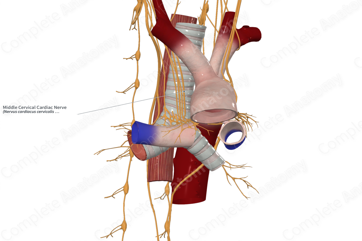
Course: Inferiorly from the middle cervical ganglion, along the posterior aspect of the common carotid artery, brachiocephalic trunk (right) or subclavian (left) artery, and along the anterior surface of the trachea to the deep cardiac plexus.
Branches: Deep cardiac plexus.
Supply: Sympathetic efferent fibers: increases heart rate, cardiac impulse conduction, and force of myocardial contraction, dilates coronary arteries.


Related parts of the anatomy
Origin
The middle cervical cardiac nerve originates in the middle cervical ganglion. Occasionally, the middle cervical ganglion is absent, and the middle cervical cardiac nerve originates directly from the sympathetic trunk in the region normally occupied by the middle cervical ganglion.
Course
Both left and right middle cervical cardiac nerves run inferiorly from the middle cervical ganglion, posterior to the common carotid artery and anterior to the longus colli muscle. The right middle cervical cardiac nerve follows the brachiocephalic artery to the anterior surface of the trachea to reach the deep cardiac plexus. The left middle cervical cardiac nerve runs down along the left common carotid artery, posterior to the subclavian artery and into the deep cardiac plexus. Some fibers from the middle cervical cardiac nerve move anteriorly, wrap around to the anterior surface of the aortic arch, and join the superficial cardiac plexus (Netter, 2011; De Gama et al., 2012).
Branches
The right and left middle cervical cardiac nerves send their fibers to the deep cardiac plexus, while some left middle cervical cardiac nerves also contribute to the superficial cardiac plexus.
Supplied Structures & Function
The middle cervical cardiac nerve carries both sympathetic efferent fibers and visceral afferent fibers.
Sympathetic efferent fibers supply the heart, and when stimulated, increase heart rate, cardiac impulse conduction, and the force of myocardial contraction, as well as dilate coronary arteries. Visceral afferent fibers from the heart, when stimulated, convey pain sensations via the superior cervical cardiac nerve towards the thoracic spinal cord.
List of Clinical Correlates
—Tachycardia
—Referred pain
References
De Gama, B. Z., Lazarus, L., Partab, P. and Satyapal, K. S. (2012) 'The Sympathetic and Parasympathetic Contributions to the Cardiac Plexus: a Fetal Study', International Journal of Morphology, 30, pp. 1569-1576.
Netter, F. H. (2011) Atlas of Human Anatomy. Netter Basic Science Series: Saunders/Elsevier.
