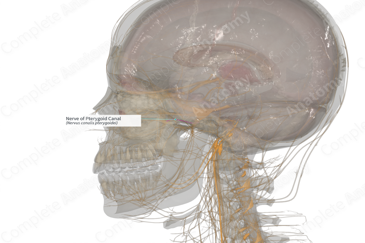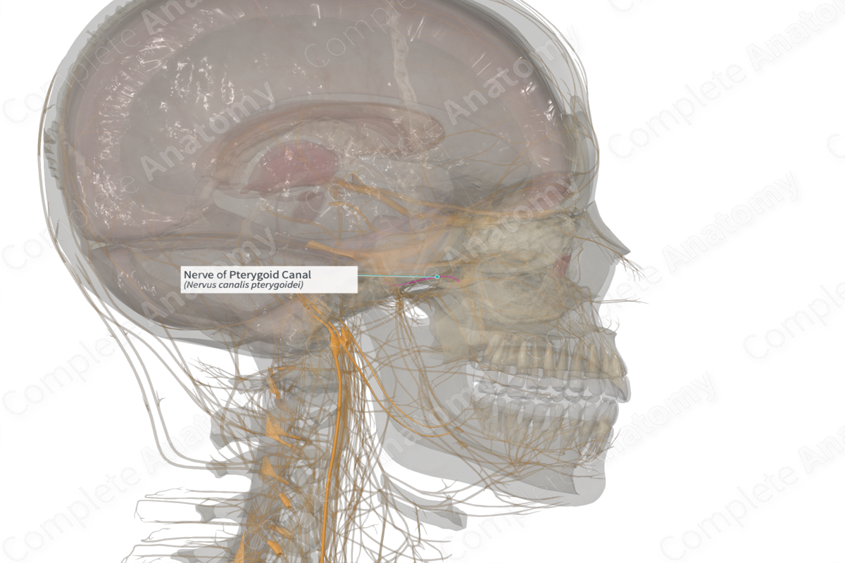
Quick Facts
Origin: Merging of the greater petrosal and deep petrosal nerves.
Course: Runs anteriorly from the foramen lacerum, through the pterygoid canal to the pterygopalatine ganglion in the pterygopalatine fossa.
Branches: None.
Supply: Sympathetic and parasympathetic innervation to the glands of the face, and sympathetic innervation to blood vessels and mucosal linings of the face.
Related parts of the anatomy
Origin
The nerve of the pterygoid canal originates when the greater petrosal nerve and the deep petrosal nerve merge in the foramen lacerum.
Course
From its origin in the foramen lacerum, the nerve of the pterygoid canal runs anteriorly into the pterygoid canal. It continues forward until it reaches the pterygopalatine fossa where it enters the pterygopalatine ganglion.
Branches
There are no named branches.
Supplied Structures
The nerve of the pterygoid canal is an autonomic nerve supplying both sympathetic and parasympathetic fibers to the mucosa, blood vessels, and glands of the head.
The portion of the nerve of the pterygoid canal derived from the greater petrosal nerve carries preganglionic parasympathetic fibers to the pterygopalatine ganglion. From here, the lacrimal gland as well as small glands in the oral cavity, nasal cavity, and palate are innervated.
The portion of the nerve of the pterygoid canal derived from the deep petrosal nerve carries postganglionic sympathetic fibers. These fibers, which originated on the superior cervical ganglion, run with the nerves associated with the pterygopalatine ganglion and the maxillary nerve to innervate blood vessels and mucosal glands of the face.
Learn more about this topic from other Elsevier products


