Description
The nerves of the shoulder region can be divided into the superficial or cutaneous and the deep nerves.
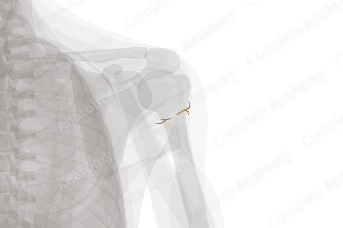
The cutaneous innervation of the shoulder region comprises the followings: supraclavicular, superior lateral brachial cutaneous, and the intercostobrachial nerve. The supraclavicular nerves are branches of the cervical plexus and contain sensory nerve fibers for the C3 and C4 cervical spinal segments. They pierce the deep fascia in the neck, descend superficial to the clavicle, and innervate the skin covering the upper half of the deltoid and the skin of the pectoral region up to the second rib. The superior lateral brachial cutaneous nerve is the distal continuation of the posterior branch of the axillary nerve. It supplies the skin covering the lower half of the deltoid. It contains sensory nerve fibers which relay information to the C5 and C6 cervical spinal segments. The intercostobrachial nerve is the lateral cutaneous branch of the second intercostal nerve. It is also related to the shoulder joint as it crosses the axilla and lies among the central group of axillary lymph nodes. The sensory dermatome distribution over the shoulder region is derived from the C3 and C4 segments of the spinal cord.
Most of the brachial plexus and its branches are destined to innervate the distal regions of the upper limb, including the arm, forearm, and hand regions; however, the brachial plexus and the proximal segments of these nerves are situated in the axillary region and are related to the shoulder in terms of the proximity of their anatomical location. The axillary nerve, which originates from the posterior cord of brachial plexus, curves back inferior to the glenohumeral articular capsule, and traverses through the quadrangular space bounded above by subscapularis and teres minor muscles. Here the axillary nerve divides into anterior and posterior branches, which provide motor innervation to deltoid and teres minor muscles (Zhao et al., 2001).
Related parts of the anatomy
References
Zhao, X., Hung, L. K., Zhang, G. M. and Lao, J. (2001) 'Applied anatomy of the axillary nerve for selective neurotization of the deltoid muscle', Clin Orthop Relat Res, (390), pp. 244-51.
Learn more about this topic from other Elsevier products


