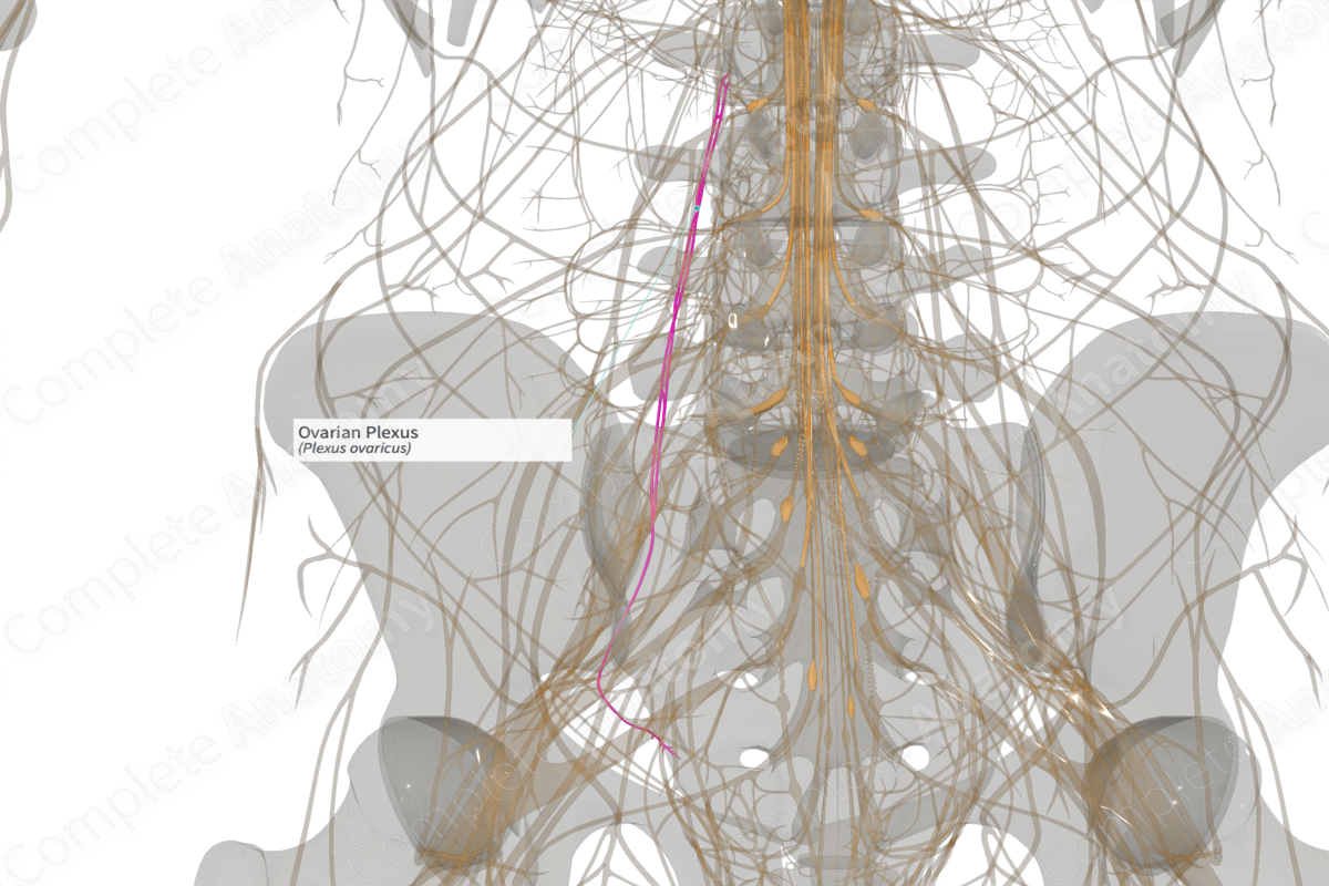
Quick Facts
Sympathetic Contribution: Greater and lesser thoracic splanchnic (T10–T11) nerves, traveling through the abdominal and prevertebral autonomic ganglia and plexuses.
Parasympathetic Contribution: Pelvic splanchnic nerves (S2-S4).
Course: Follows the course of the ovarian artery to reach the pelvis.
Sympathetic Supply: Ovary, distal uterine tube, and uterine fundus.
Parasympathetic Supply: Ovary, distal uterine tube, and uterine fundus.


Contributing Nerves
Sympathetic nerves descend with the ovarian artery in a fold of peritoneum that forms the suspensory ligament of the ovary. Some fibers pass through the superior and inferior hypogastric plexuses. Parasympathetic fibers cross the pelvic floor to the inferior hypogastric and ovarian plexuses.
Course
Descending sympathetic fibers work their way down the posterior abdominal wall in aortic, renal, periarterial, and ureteric plexuses. Those from the latter two plexuses follow the ovarian artery within the suspensory ligament of the ovary. Ascending sacral and pelvic splanchnic nerves pass along the pelvic floor. Some fibers ascend within the broad ligament with the uterine artery. Others continue medially to the inferior and superior hypogastric plexuses.
Branches
No named branches come from the ovarian plexus.
Supplied Structures
Ovary, fundus of the uterus, and distal uterine tube.
