Quick Facts
Sympathetic Contribution: T6-L2 spinal cord sympathetic neurons; greater splanchnic nerve; celiac ganglion postsynaptic sympathetic neurons.
Parasympathetic Contribution: Vagal parasympathetic axons.
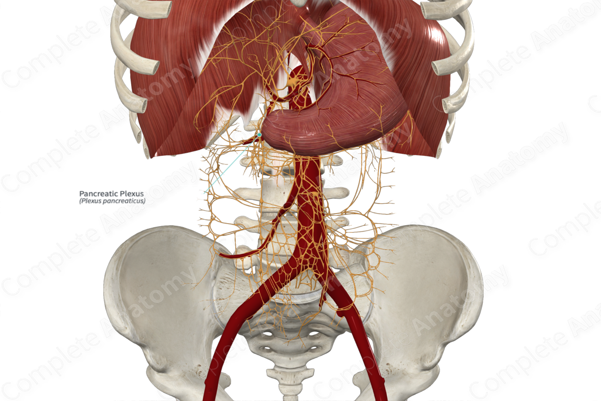
Course: Lies on the posterosuperior portion of the head of the pancreas.
Sympathetic Supply: Pancreatic islets, vasculature, ducts, and acini.
Parasympathetic Supply: Innervates both endocrine and exocrine cells in the pancreas.


Contributing Nerves
The pancreatic plexus is a subdivision of the celiac plexus and the posterior hepatic plexus. It consists of sympathetic axons from the celiac ganglion and parasympathetic axons from the posterior vagal trunk. Visceral sensory axons from the pancreas travel through the pancreatic plexus to reach the central nervous system (Babic and Travagli, 2016).
Course
The pancreatic plexus is primarily visible in the posterosuperior portion of the head of the pancreas (Ren et al., 2020). Smaller fascicles of axons spread out to innervate more distal pancreatic tissue but are not visible.
Branches
There are no named branches.
Supplied Structures
The pancreatic plexus conveys all efferent axons to the pancreas, innervating pancreatic islets, acini, ducts, and vasculature. Both sympathetic and parasympathetic innervation affects endocrine function, while parasympathetic efferent fibers are the primary source of innervation to exocrine acini (Babic and Travagli, 2016).
Visceral sensory fibers carrying afferents from the pancreas pass through the pancreatic plexus.
References
Babic, T. and Travagli, R. A. (2016) 'Neural Control of the Pancreas.'. DOI: 10.3998/panc.2016.27.
Ren, K., Yi, S.-Q., Dai, Y., Kurosawa, K., Miwa, Y. and Sato, I. (2020) 'Clinical anatomy of the anterior and posterior hepatic plexuses, including relations with the pancreatic plexus: A cadaver study', Clinical Anatomy, 33(5), pp. 630-636.
Learn more about this topic from other Elsevier products

