Description
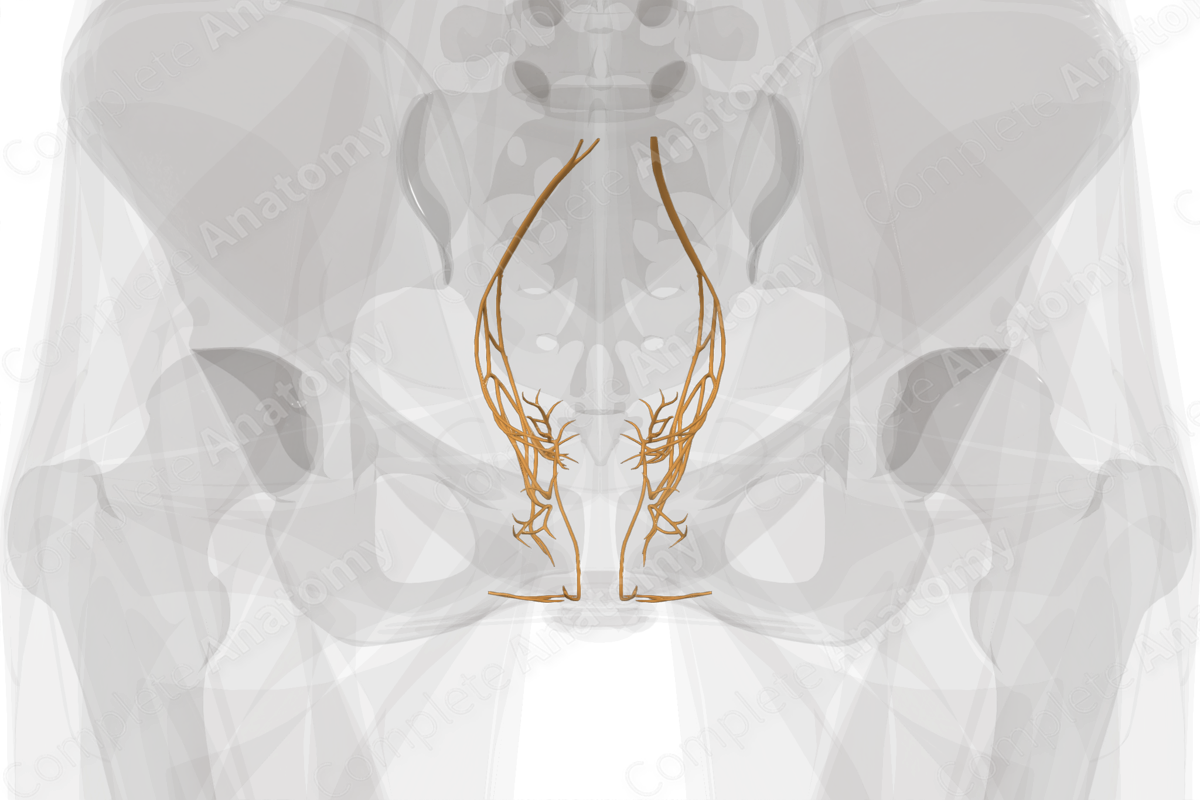
The pelvic plexuses are weblike collections of autonomic nerve axons found near the pelvic viscera in the pelvic cavity. Unlike the abdominal plexuses which typically have identifiable ganglia, the pelvic plexuses have smaller more discrete ganglia which, unique among all autonomic ganglia, contain both sympathetic and parasympathetic neuronal cell bodies (Dail, 1996).
The inferior hypogastric plexus forms the bulk of the pelvic plexuses and is a large network of fibers and diffuse ganglia lying bilaterally just medial and anterior to the internal iliac artery and external to the peritoneum. It receives sympathetic fibers from sacral splanchnic nerves of the sympathetic chain, or from the aortic plexus via the hypogastric nerve, which connects the superior and inferior hypogastric plexuses. Parasympathetic fibers from S2-S4 travel to the inferior hypogastric plexus via the pelvic splanchnic nerves. Functionally, the pelvic plexuses serve the tissues of the middle rectum, pelvic viscera, bladder, and reproductive organs.
Extending from the inferior hypogastric plexus are the middle and inferior rectal plexuses, the vesical plexus, the deferential and prostatic plexuses in males, and the uterovaginal plexus in females. These convey autonomic innervation to the middle portion of the rectum or to the bladder and reproductive organs (Standring, 2016).
References
Dail, W. G. (1996) 'The pelvic plexus: Innervation of pelvic and extrapelvic visceral tissues', Microscopy Research and Technique, 35(2), pp. 95-106.
Standring, S. (2016) Gray's Anatomy: The Anatomical Basis of Clinical Practice. Gray's Anatomy Series 41 edn.: Elsevier Limited.


