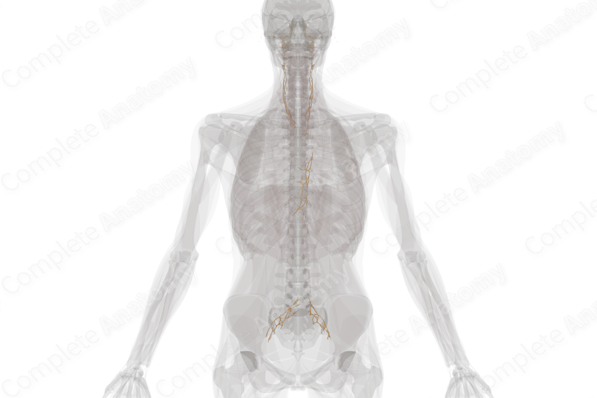
Description
The perivascular plexuses are collections of primarily postganglionic sympathetic axons that follow the vasculature. All arteries, as well as some larger veins and lymph vessels, have sympathetic axons that travel along their surface (Standring, 2016), both innervating the smooth muscle of the vessel wall and following it to targets in the distal arterial territory. Any collection of sympathetic axons traveling along an artery can be considered a perivascular plexus.
Some major examples of perivascular plexuses are as follows.
—In the head, all postganglionic sympathetic axons ascend from the cervical ganglia up along the common carotid, external carotid, and internal carotid arteries. They do so as carotid plexuses named after the particular artery along which they are found.
—In the thorax and abdomen, the aorta is covered by a web of sympathetic axons that forms the aortic plexus. In the lower abdomen when the aorta bifurcates, the aortic plexus also splits to form the iliac plexuses.


References
Standring, S. (2016) Gray's Anatomy: The Anatomical Basis of Clinical Practice. Gray's Anatomy Series 41 edn.: Elsevier Limited.
Learn more about this topic from other Elsevier products
Plexus

Visceral plexuses are a network of nerve fiber and ganglia surrounding organs of the abdomen and pelvis region that convey sympathetic, parasympathetic, and visceral afferent input.
