Quick Facts
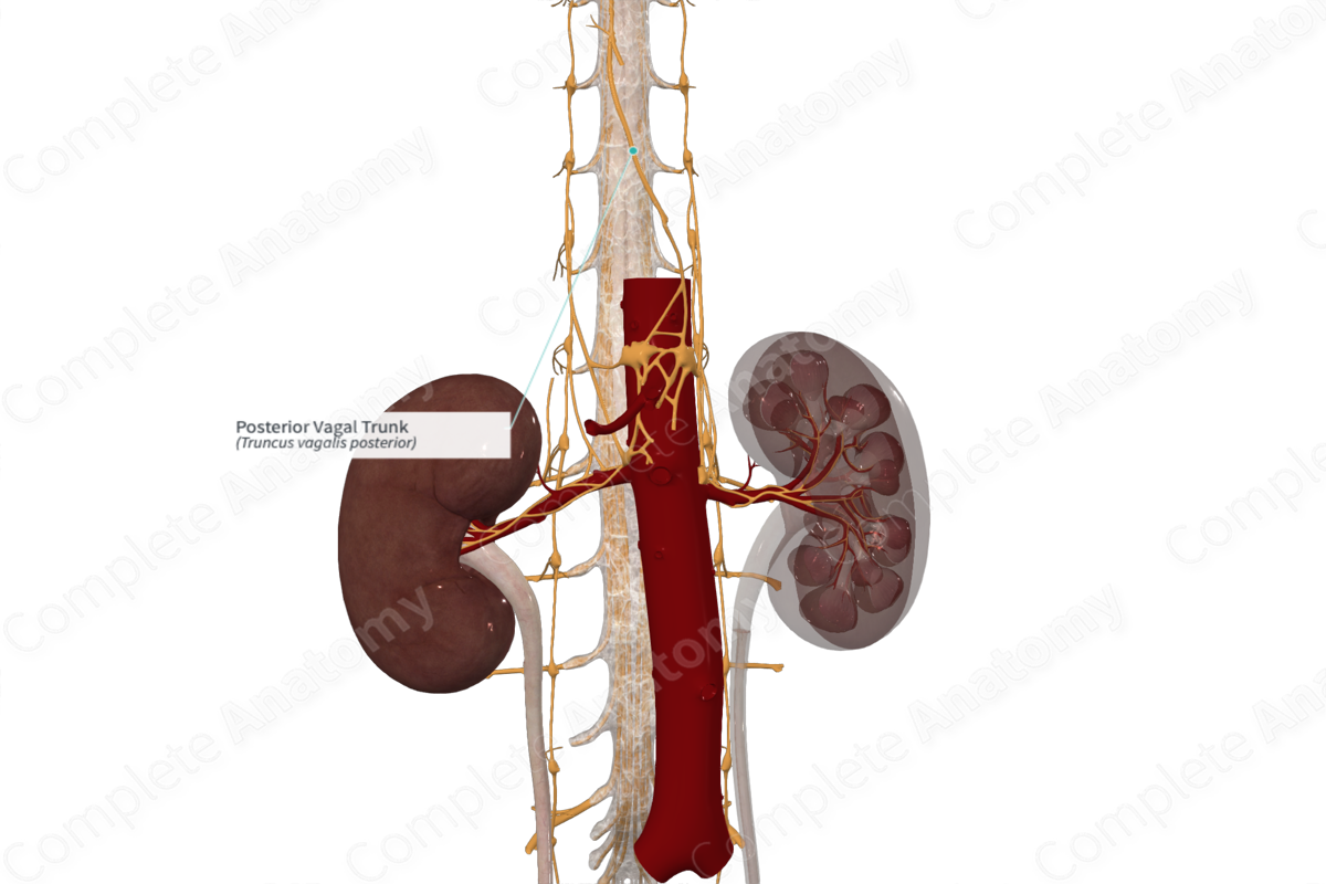
Origin: Formed by coalescence of parasympathetic fibers of the posterior esophageal plexus, just superior to the diaphragm.
Course: Passes along the posterior surface of the esophagus, through the esophageal hiatus of the diaphragm into the abdomen.
Branches: Gastric (posterior) and celiac branches.
Supply: Mixed nerve supplying visceral sensory and parasympathetic innervation to the foregut and midgut.
Related parts of the anatomy
Origin
After passing posterior to the root of the right lung, the right vagus nerve passes inferomedially and posteriorly, and branches to form the posterior esophageal plexus. At the distal end of the esophagus, just prior to its passage through the diaphragm, the remaining axons coalesce to form the posterior vagal trunk.
Course
The posterior vagal trunk accompanies the esophagus. It passes between the posterior surface and posterior rim of the esophageal hiatus of the diaphragm to enter the abdominal cavity.
Branches
Upon entering the abdominal cavity, the posterior vagal trunk gives off short posterior gastric nerves that travel posteriorly. However, the majority of the axons of the posterior vagal trunk travel to the celiac trunk, renal, and superior mesenteric arteries. They form periarterial plexuses around these vessels and follow them out to their targets.
Supplied Structures & Function
The posterior vagal trunk is a mixed nerve with both visceral sensory and parasympathetic fibers. It supplies the posteroinferior portion and greater curvature of the stomach, and spleen. Fibers pass through a number of abdominal autonomic ganglia, including the celiac, aorticorenal, and superior mesenteric ganglia. These distribute parasympathetic preganglionic axons throughout the foregut and midgut.


