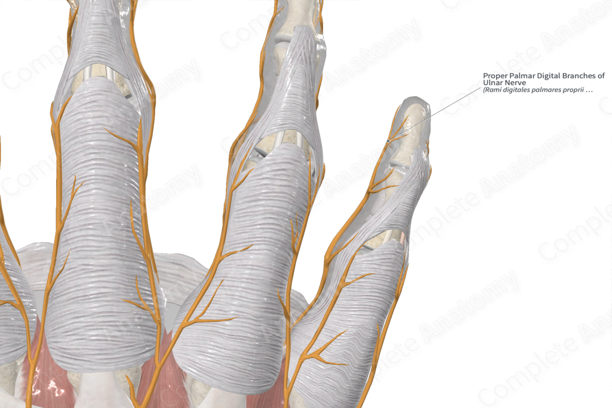
Proper Palmar Digital Branches of Ulnar Nerve
Rami digitales palmares proprii nervi ulnaris
Read moreQuick Facts
Origin: Common palmar digital nerves of ulnar nerve (or directly from the superficial terminal branch of ulnar nerve; C8-T1).
Course: Travel through the digits with their corresponding digital arteries and veins.
Branches: None.
Supply: Skin of the medial one and a half digits (little finger and medial half of the ring finger).


Related parts of the anatomy
Origin
The proper palmar digital nerve for the medial side of the little finger arises directly from the superficial terminal branch of ulnar nerve. Those for the interdigital skin between the adjacent side of the little and ring fingers arise from the common palmar digital branches of the ulnar nerve.
Course
The ulnar nerve enters the palm by passing superficial to the flexor retinaculum and lateral to pisiform bone, while being covered by a superficial slip of the retinaculum (volar carpal ligament). At the distal border of retinaculum, the nerve terminates by dividing into its superficial and deep branches.
Just distal to the pisiform bone, the superficial branch gives off two palmar digital branches. The medial of the two branches is a proper palmar digital nerve which innervates the skin of the medial side of the little finger. The lateral branch is a common palmar digital nerve, which further divides into two proper palmar digital nerves. These innervate the skin of adjacent sides of the ring and little fingers. It also sends a communicating branch to the palmar digital nerves of the median nerve.
Branches
Sensory nerve fibers through the proper palmar digital branches of the ulnar nerve eventually end up relaying sensory information into the C8 & T1 cervical and thoracic segments of the spinal cord.
Supplied Structures
Cutaneous sensory innervation to the skin of the medial one and a half digits (little finger and medial half of the ring finger).
Learn more about this topic from other Elsevier products

