Quick Facts
Sympathetic Contribution: Thoracic pulmonary branches of thoracic ganglia.
Parasympathetic Contribution: Vagus nerve, recurrent laryngeal nerve, and bronchial branches of vagus nerve.
Course: Surrounds the pulmonary arteries and main bronchi.
Sympathetic Supply: Contraction of pulmonary and bronchial vasculature, relaxes smooth muscle of airways, and inhibits mucus secretion.
Parasympathetic Supply: Contraction of bronchial smooth muscle, relaxes pulmonary and bronchial vasculature, and increases secretion of mucus.


Related parts of the anatomy
Contributing Nerves
The nerves of the pulmonary plexus originate from the thoracic branches of the upper sympathetic trunk (sympathetic contribution) and the vagus nerve (parasympathetic contribution). For both sympathetic and parasympathetic fibers, axons may travel directly to the pulmonary plexus, or may travel through the cardiac plexus first.
Course
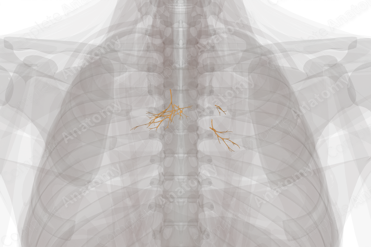
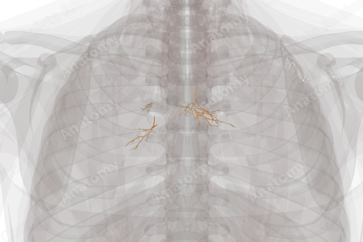
The pulmonary plexus is the plexus that surrounds the roots of each lung. It is contiguous with and lateral to the deep cardiac plexus.
—The left pulmonary plexus surrounds the left primary bronchus and the left pulmonary artery at the root of the left lung.
—The right pulmonary plexus surrounds the right primary bronchus and the right pulmonary artery at the root of the right lung.
Both right and left plexuses can be arbitrarily divided into anterior and posterior plexuses, corresponding to fibers located on the anterior or posterior root of the lung, respectively.
—The anterior pulmonary plexus receives only a few anterior bronchial branches from the vagus nerve. These combine with sympathetic fibers in front of the root of the lung.
—The posterior pulmonary plexus is larger and receives many posterior bronchial branches from the vagus nerve. These combine with sympathetic fibers posterior to the root of the lung.
Branches
The pulmonary plexus can be divided into four different plexuses:
—the right anterior pulmonary plexus;
—the right posterior pulmonary plexus;
—the left anterior pulmonary plexus;
—the left posterior pulmonary plexus.
Fibers that leave the plexus travel laterally to the lung tissue by following the bronchopulmonary tree, splitting as the bronchi and pulmonary vasculature splits (Kaminsky, 2011).
Supplied Structures
The pulmonary plexus provides both sympathetic and parasympathetic innervation to the lungs and carries visceral afferents back to the CNS.
Sympathetic efferents in the pulmonary plexus are thought to cause contraction of pulmonary and bronchial vasculature, relax smooth muscle of the airways, and inhibit secretion of mucus from the epithelial linings of the lung. Parasympathetic efferents in the pulmonary plexus cause either contraction or relaxation of the smooth muscle of the airways, although it is typically associated with contraction of bronchi. Additional functions include relaxation of pulmonary and bronchial vasculature, as well as increased secretion of mucus from the epithelial lining of the lungs (Chila, 2010).
Visceral afferents travel from the lungs back via both sympathetic nerves to the spinal cord, and the vagal nerves to the brainstem. The functions of spinal afferents are largely unknown but may include pain sensation. The functions of vagal afferents include mediating protective reflexes such as cough and mucus secretion, lung stretch as measured by stretch receptors in the alveoli, and pain sensation (Mazzone and Canning, 2013).
List of Clinical Correlates
—Asthma
—COPD
—Pulmonary hypertension
—Pulmonary embolism
—Neurogenic pulmonary edema
References
Chila, A. G. (2010) Foundations of Osteopathic Medicine. 3rd edn.: Lippincott Williams & Wilkins.
Kaminsky, D. (2011) Netter Collection of Medical Illustrations: Respiratory System E-Book. Netter Green Book Collection: Elsevier Health Sciences.
Learn more about this topic from other Elsevier products

