Quick Facts
Sympathetic Contribution: Least thoracic splanchnic nerve via the aorticorenal ganglion, in addition to the lesser thoracic and first lumbar splanchnic nerves.
Parasympathetic Contribution: Branches from the vagal trunks.
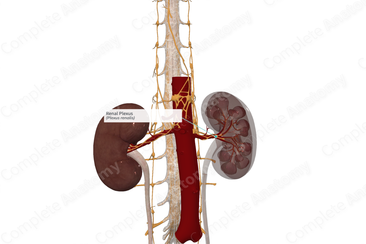
Course: Forms around and follows the renal artery.
Sympathetic Supply: Kidney and suprarenal gland, via branches to the ureter and gonads.
Parasympathetic Supply: Ureter and gonads.


Contributing Nerves
Preganglionic sympathetic fibers travel primarily in the least thoracic splanchnic nerve, with variable contributions from the lesser thoracic and first lumbar splanchnic nerves. These synapse primarily on the aorticorenal ganglion but may also include the celiac and/or superior mesenteric ganglia. Preganglionic parasympathetic nerves of vagal origin join the renal plexus.
Course
The renal plexus follows the course of the renal artery to the renal medulla.
Branches
Before entering the renal hilum, branches peel off the renal plexus to surround the ureteric and gonadal arteries. These form plexuses of the same name. In addition, other axons follow the inferior suprarenal arteries providing innervation to the suprarenal gland.
Supplied Structures
The renal plexus supplies the blood vessels, renal glomeruli, juxtaglomerular apparatus, and tubules of the kidney. They control renal blood flow, renin release, and sodium reabsorption (Shannon et al., 1998).
References
Shannon, J. L., Headland, R., MacIver, A. G., Ferryman, S. R., Barber, P. C. and Howie, A. J. (1998) 'Studies on the innervation of human renal allografts', J Pathol, 186(1), pp. 109-15.
Learn more about this topic from other Elsevier products

