Description
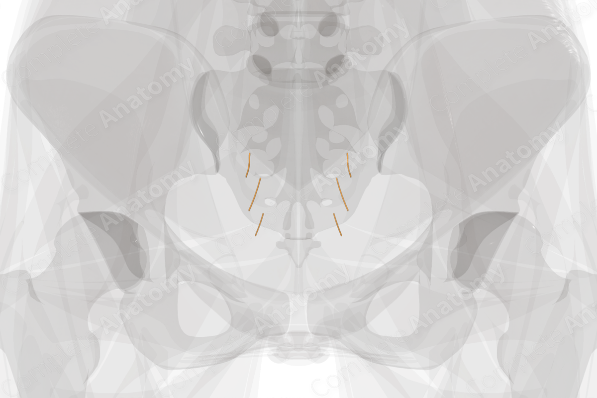
The sacral part of the autonomic division refers to the parasympathetic fibers that originate in the lateral horn at the S2-S4 spinal cord levels. These fibers form pelvic splanchnic nerves which run to the hypogastric plexuses and from there to glands, smooth muscle, and viscera of the hindgut, bladder, and reproductive organs.
In the hindgut, sacral parasympathetic fibers drive smooth muscle contraction to propel fecal matter towards the anus, as well as glandular secretion (Standring, 2016).
In the bladder, sacral parasympathetic fibers drive smooth muscle contraction to empty the bladder.
In the pelvis, sacral parasympathetic fibers are vasodilators of the erectile tissue of the penis and clitoris.


Related parts of the anatomy
References
Standring, S. (2016) Gray's Anatomy: The Anatomical Basis of Clinical Practice. Gray's Anatomy Series 41 edn.: Elsevier Limited.
Learn more about this topic from other Elsevier products
Autonomic Function

Respiration is a fundamental autonomic function that involves the coordination of the diaphragm activity, sensors for lung volume and blood gases, modulation of airway tone, and control of the tongue continuously over the duration of one’s life.
