Quick Facts
Origin: Fourth lumbar to third sacral nerves (L4—S3).
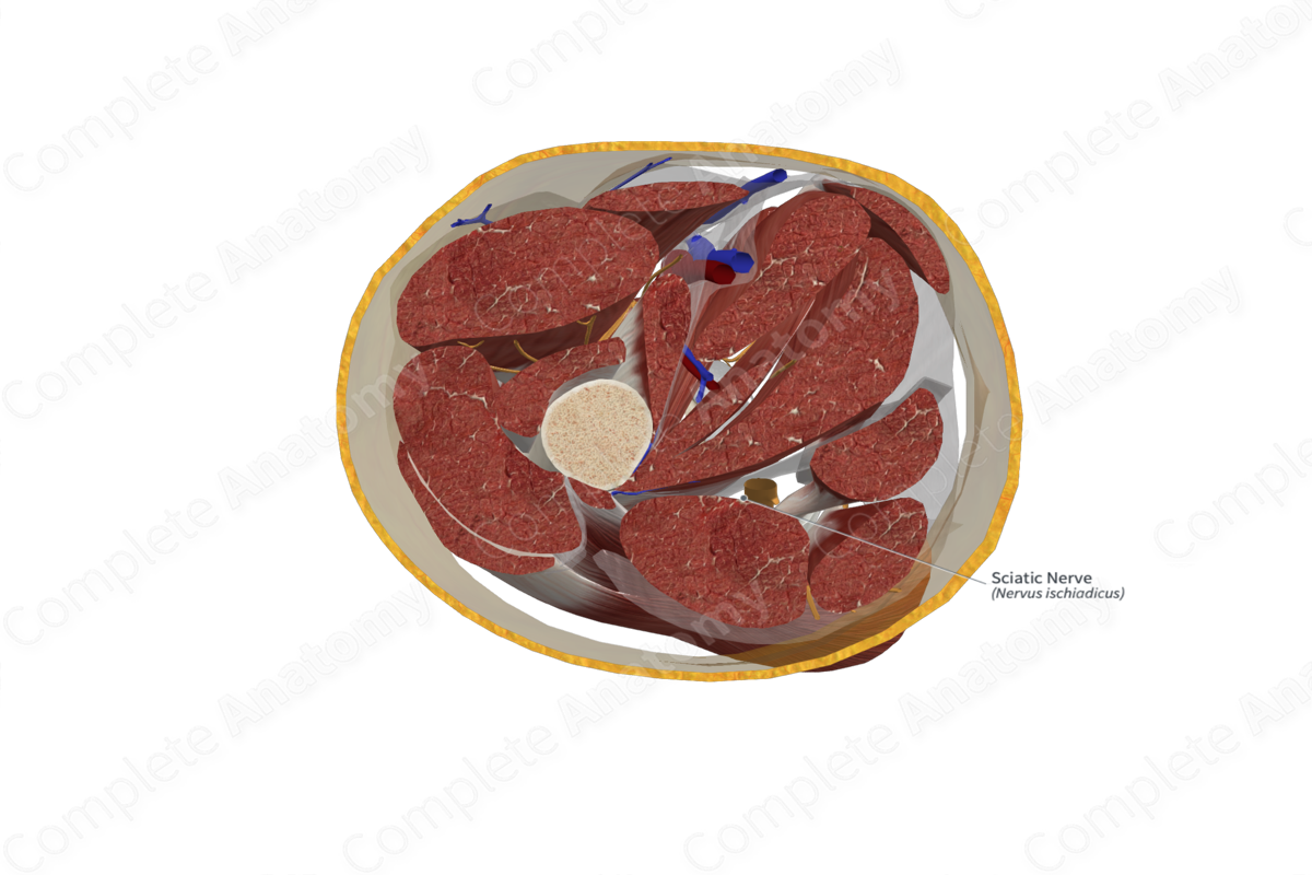
Course: Enters the gluteal region through the greater sciatic foramen, beneath the piriformis muscle. It descends over the lateral rotators of the hip. It passes through the posterior compartment of the thigh to reach the superior border of the popliteal fossa.
Branches: Muscular branches and the tibial and common fibular nerve.
Supply: Motor innervation to the posterior compartment of the thigh via muscular branches; the posterior compartment of the leg via the tibial nerve; the muscles of the anterior and lateral compartments of the leg via the common fibular nerve. Sensory innervation to the skin of the leg and foot via the common fibular and tibial nerves.
Related parts of the anatomy
Origin
The sciatic nerve arises from the anterior rami of the fourth lumbar to third sacral nerves (L4—S3).
Course
The sciatic nerve enters the gluteal region through the greater sciatic foramen beneath the piriformis. It descends over obturator internus, gemelli, and quadratus femoris, between the ischial tuberosity and the greater trochanter. It descends in the posterior compartment of the thigh until it reaches the superior border of the popliteal fossa. Here, it divides into tibial and common fibular nerves (Moore, Dalley and Agur, 2013; Adibatti and Sangeetha, 2014).
Branches
In the thigh, the sciatic nerve gives off muscular branches to supply the hamstring muscles. Its terminal bifurcation gives rise to the tibial and common fibular nerves, which descend into the leg.
Supplied Structures
In the thigh, the muscular branches of the sciatic nerve supply the semimembranosus, semitendinosus, biceps femoris, and adductor magnus muscles.
The tibial nerve supplies branches to the superficial and deep muscles in the posterior compartment of the leg (gastrocnemius, soleus, plantaris, tibialis posterior, flexor digitorum longus, and flexor hallucis longus). It gives off the sural nerve (which supplies cutaneous innervation to the skin on lateral part of the back of the leg), and a recurrent articular branch (which supplies the knee joint).
The superficial fibular nerve innervates peroneus longus and brevis. It pierces the deep fascia to innervate the skin on the lateral side of the lower leg and the dorsum of the foot. The deep fibular nerve provides motor innervation to the muscles in the extensor compartment of the leg and becomes sensory cutaneous for the skin in the first interdigital space.
List of Clinical Correlates
—Sciatica
—Piriformis syndrome
—Injury to sciatic nerve
—Foot drop
References
Adibatti, M. and Sangeetha, V. (2014) 'Study on variant anatomy of sciatic nerve', J Clin Diagn Res, 8(8), pp. AC07-9.
Moore, K. L., Dalley, A. F. and Agur, A. M. R. (2013) Clinically Oriented Anatomy. Clinically Oriented Anatomy 7th edn.: Wolters Kluwer Health/Lippincott Williams & Wilkins.


