Quick Facts
Origin: Inner walls of the ventricles, deep to the endocardium.
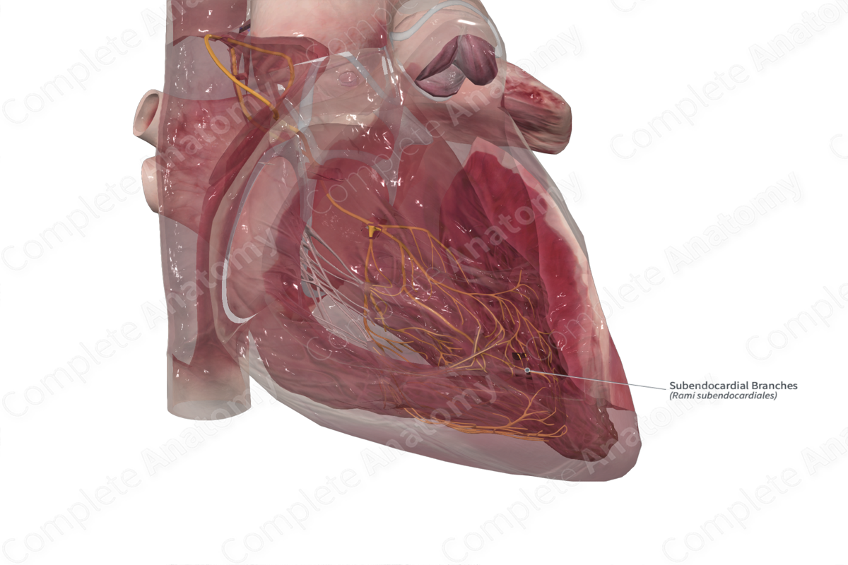
Course: Spread throughout the subendocardium of the ventricles.
Branches: None.
Supply: Transmit an action potential to the myocardial fibers of the ventricles.


Origin
The subendocardial branches (or Purkinje fibers) are the terminal branches of the atrioventricular bundle.
Course
The Purkinje fibers form the last portion of the unidirectional cardiac conduction system. They are found spread throughout the entire inner surface of the ventricles, just deep to the endocardium in a space called the subendocardium.
Branches
There are no branches for the Purkinje fibers. They connect directly to the myocardial fibers.
Supplied Structures & Function
Preceding a ventricular contraction, the Purkinje cells receive the depolarizing action potential from the left and right bundle of the AV bundle. The fast conducting Purkinje fibers spread the action potential rapidly to the entire inner surface of the ventricles, enabling synchronization of ventricular contraction. The action potential in the Purkinje fibers first acts on the adjacent myofibers of the inner ventricular myocardium. This contraction then travels through the myocardium, spreading outward until it reaches the outer ventricular walls under the epicardium (Katz, 2010, Anderson et al., 2009).
References
Anderson, R. H., Yanni, J., Boyett, M. R., Chandler, N. J. and Dobrzynski, H. (2009) 'The anatomy of the cardiac conduction system', Clin Anat, 22(1), pp. 99-113.
Katz, A. M. (2010) Physiology of the Heart. M - Medicine Series: Wolters Kluwer Health/Lippincott Williams & Wilkins Health.
Learn more about this topic from other Elsevier products
Purkinje Fiber

The cardiac conducting system, also known as the cardiac electric system, consists of Purkinje fibers, the bundle of His, the atrioventricular node (AVN), and the sinoatrial node (SAN) [13].
