Quick Facts
Sympathetic Contribution: Lumbar and sacral splanchnic nerves.
Parasympathetic Contribution: Pelvic splanchnic nerves.
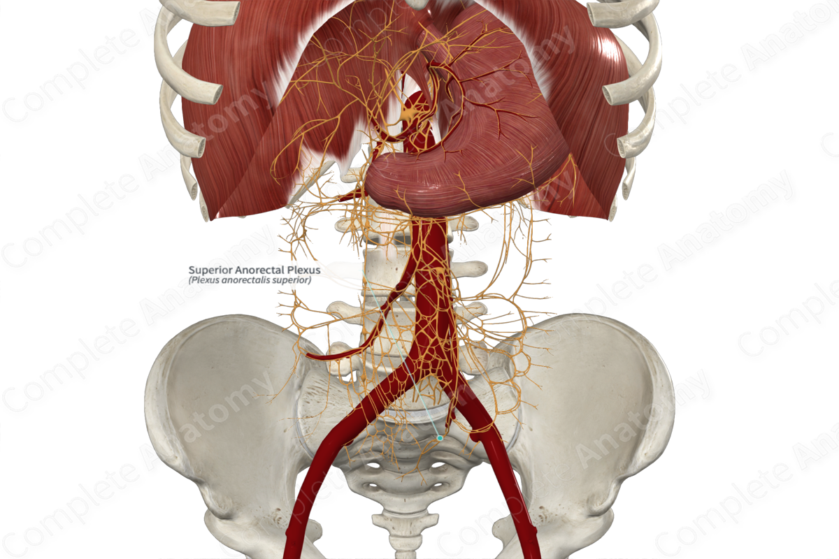
Course: Follows the superior rectal branch of the inferior mesenteric artery inferiorly to the rectum.
Sympathetic Supply: Mainly to vasculature in the wall of the rectum.
Parasympathetic Supply: Mainly to goblet cells in the rectal epithelium, smooth muscle coat of the rectal wall, and the internal anal sphincter.


Contributing Nerves
The sympathetic component of the superior anorectal plexus arises from two sources. The first are from lumbar splanchnic nerves that terminate in the inferior mesenteric and aortic plexuses. The second are from sacral splanchnic nerves (preganglionic axons that arise from first and second lumbar levels L1 and L2) but descend in the sympathetic trunk to the second to fourth sacral levels (S2-S4).
The parasympathetic contribution to the superior anorectal plexus arises from second to fourth sacral spinal cord levels (S2—S4) and unite to form pelvic splanchnic nerves. Pelvic and sacral splanchnic axons travel together to the inferior anorectal plexus. They subsequently pass into the hypogastric nerve and superior hypogastric plexus. Sympathetic fibers terminate on ganglia in these plexuses. From any of these sites, they pass towards the superior anorectal artery and contribute to the superior anorectal plexus surrounding it.
Course
Usually pelvic splanchnic (parasympathetic) nerves pass to the inferior hypogastric plexus and ascend within or along the hypogastric nerve to the superior hypogastric plexus. Here, they contribute to the inferior anorectal plexus formed around the inferior anorectal artery. Some pelvic splanchnic nerves, upon reaching the superior hypogastric plexus, pass directly to the distal superior anorectal artery and join the plexus. Other nerves pass directly to the superior rectum.
Branches
No named branches arise from the superior anorectal plexus.
Supplied Structures
The superior anorectal plexus supplies the glandular cells of the rectal epithelium, superficial smooth muscle of the muscularis layer, and vasculature of the superior third of the rectum. It controls blood flow (primarily sympathetic), mucous secretion, and contraction (both, primarily parasympathetic) of the superior part of the rectum.
Visceral sensory fibers carrying afferents from the rectum pass through the superior mesenteric plexus.
List of Clinical Correlates
—Colonic propulsion and defecation
—Relaxation of the internal anal sphincter
Learn more about this topic from other Elsevier products
Plexus

Visceral plexuses are a network of nerve fiber and ganglia surrounding organs of the abdomen and pelvis region that convey sympathetic, parasympathetic, and visceral afferent input.
