Description
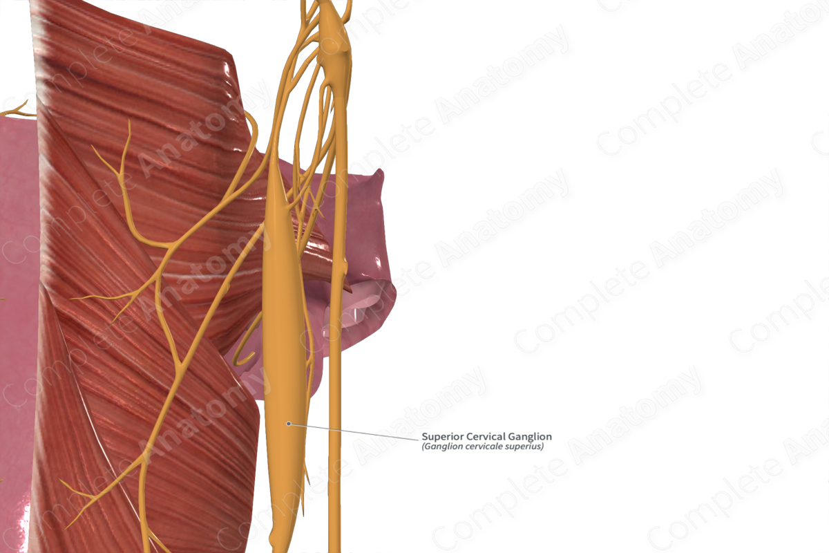
The superior cervical ganglion is the largest of the cervical ganglia and lies inferior to the occiput and anterior to the transverse processes of the second and third cervical vertebrae, in front of the longus capitis muscle (Civelek et al., 2008). It is related anteriorly to the internal carotid artery. Inferiorly, the ganglion connects with the middle cervical ganglion via an interganglionic branch.
The cervical sympathetic trunk is an extension of the thoracic sympathetic trunk, receiving its preganglionic fibers from it. These preganglionic sympathetic fibers originate from the lateral gray horn of the T1 spinal segment and enter the sympathetic trunk via the white rami communicans of the first thoracic nerve. Within the trunk, the preganglionic neurons ascend to synapse with the cell bodies of the postganglionic neurons inside the superior cervical ganglion. From here onwards, the postganglionic sympathetic fibers are distributed throughout the head and neck following various routes.
—Some postganglionic fibers form a plexus around the common, internal, and external carotid arteries. From here, the sympathetic neurons are distributed to various parts of the head and neck, tracing the course of the arterial branches.
—A large proportion of the postganglionic sympathetic fibers enter the anterior rami of first four cervical nerves (C1—C4) via the gray rami communicantes. In addition, a few gray rami communicantes are also given to structures, including the inferior vagal ganglion, hypoglossal nerve, superior jugular bulb, meninges, and to the inferior glossopharyngeal and vagal ganglia (via the jugular nerve).
—A few medial branches from the superior cervical ganglion are distributed to the carotid body, pharyngeal plexus, and to the heart via superior cervical cardiac nerve.
List of Clinical Correlates
—Stellate ganglion block
—Horner’s syndrome
References
Civelek, E., Karasu, A., Cansever, T., Hepgul, K., Kiris, T., Sabanci, A. & Canbolat, A. (2008) Surgical anatomy of the cervical sympathetic trunk during anterolateral approach to cervical spine. European spine journal : official publication of the European Spine Society, the European Spinal Deformity Society, and the European Section of the Cervical Spine Research Society, 17(8), 991-995.
Learn more about this topic from other Elsevier products


