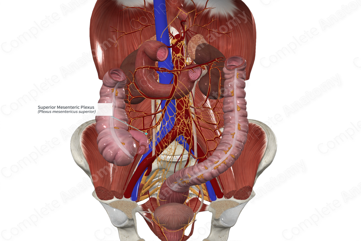
Quick Facts
Sympathetic Contribution: Lesser thoracic splanchnic nerves (T10-T11).
Parasympathetic Contribution: Vagus nerves.
Course: Follows the vagus nerve.
Sympathetic Supply: Midgut.
Parasympathetic Supply: To intramural parasympathetic ganglia located in the wall of organs and glands of midgut.
Contributing Nerves
Preganglionic nerves arise from tenth and eleventh thoracic nerves (T10—T11) and form the lesser splanchnic nerve. These terminate primarily in the superior mesenteric ganglion. Postganglionic axons form the superior mesenteric plexus following the superior mesenteric artery and its branches. These are joined by preganglionic parasympathetic nerves from the vagal trunks.
Course
The superior mesenteric plexus follows the superior mesenteric artery, passing between layers of peritoneum that form the mesentery, to reach components of the midgut.
Branches
The superior mesenteric plexus follows branches of the superior mesenteric artery and forms a number of secondary plexuses, including the inferior pancreaticoduodenal, intestinal, ileocolic, and right and middle colic plexuses.
Supplied Structures
The superior mesenteric plexus contributes autonomic innervation to the vascular territory of the superior mesenteric artery. This includes the distal portion of the duodenum, inferior pancreas, jejunum, ileum, cecum, appendix, ascending colon, and the proximal half to two thirds of the transverse colon.
Visceral sensory fibers carrying afferents from the aforementioned organs pass through the superior mesenteric plexus.
List of Clinical Correlates
—Superior mesenteric artery syndrome


